当前位置:
X-MOL 学术
›
J. Alloys Compd.
›
论文详情
Our official English website, www.x-mol.net, welcomes your
feedback! (Note: you will need to create a separate account there.)
Structure and optical properties of La2-Gd SiO5:Dy3+ phosphors
Journal of Alloys and Compounds ( IF 5.8 ) Pub Date : 2019-02-01 , DOI: 10.1016/j.jallcom.2018.10.090 Simon N. Ogugua , Samy K.K. Shaat , Hendrik C. Swart , Robin E. Kroon , Odireleng M. Ntwaeaborwa
Journal of Alloys and Compounds ( IF 5.8 ) Pub Date : 2019-02-01 , DOI: 10.1016/j.jallcom.2018.10.090 Simon N. Ogugua , Samy K.K. Shaat , Hendrik C. Swart , Robin E. Kroon , Odireleng M. Ntwaeaborwa
|
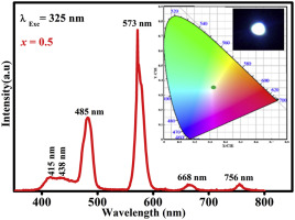
Abstract Lanthanum gadolinium oxyorthosilicate or La2-xGdxSiO5 (x = 0, 0.5, 1.0, 1.5 and 2.0) nanophosphors doped with dysprosium (Dy3+) were prepared by urea- and ammonium nitrate-assisted solution combustion method. The X-ray diffraction (XRD) patterns confirmed that the phosphors crystallized in a mixed phase of La2SiO5 and La(OH)3 and a pure monoclinic phase of Gd2SiO5 or the admixtures of the three phases depending on the ratio of La:Gd in the host lattice. The estimated crystallite sizes were found to vary from 10 to 21 nm. The field emission scanning electron microscopy (FE-SEM) images showed that the particles were agglomerated together and they had no definite sizes. The chemical composition analyses and the electronic states were analyzed using the energy-dispersive X-ray spectroscopy (EDS) and X-ray photoelectron spectroscopy (XPS) respectively. The Fourier transform infrared spectroscopy (FTIR) data supplemented both the XRD and EDS data by confirming that the stretching mode frequencies were all related to La2SiO5 and Gd2SiO5, except a few absorption peaks ascribed to atmospheric moisture and hydrocarbons. The band gaps measured from the ultraviolet visible spectroscopy (UV–Vis) data were shown to vary with the molar ratio of La to Gd. The photoluminescence spectra showed two characteristic emissions of Dy3+ at 485 nm (blue) and 573 nm (yellow) and an additional broad emission (in the blue region) with a maximum at ∼415 nm. The International Commission on Illumination (CIE) chromaticity coordinates calculated from the fluorescence emission showed colours which were tuned from blue to white and yellow when the molar ratio of La to Gd in the La2-xGdxSiO5:Dy3+ lattice was varied. Depending on the excitation wavelength, energy transfer was observed from Dy3+ substituted in Gd3+ lattice sites to Dy3+ substituted in La3+ lattice sites. The internal photoluminescence quantum yield of the phosphors was measured using an integrating sphere method.
中文翻译:

La2-Gd SiO5:Dy3+荧光粉的结构和光学性质
摘要 采用尿素和硝酸铵辅助溶液燃烧法制备了掺杂镝(Dy3+)的镧钆氧正硅酸盐或La2-xGdxSiO5 (x = 0, 0.5, 1.0, 1.5 and 2.0)纳米磷光体。X 射线衍射 (XRD) 图案证实荧光粉在 La2SiO5 和 La(OH)3 的混合相和 Gd2SiO5 的纯单斜相或三相的混合物中结晶,这取决于 La2SiO5 和 La(OH)3 的比例。主晶格。发现估计的微晶尺寸从 10 到 21 nm 不等。场发射扫描电子显微镜 (FE-SEM) 图像显示颗粒聚集在一起并且它们没有确定的尺寸。分别使用能量色散 X 射线光谱 (EDS) 和 X 射线光电子能谱 (XPS) 分析化学成分分析和电子态。傅里叶变换红外光谱 (FTIR) 数据通过确认拉伸模式频率都与 La2SiO5 和 Gd2SiO5 相关,除了少数归因于大气水分和碳氢化合物的吸收峰外,补充了 XRD 和 EDS 数据。从紫外可见光谱 (UV-Vis) 数据测量的带隙显示出随 La 与 Gd 的摩尔比而变化。光致发光光谱显示 Dy3+ 在 485 nm(蓝色)和 573 nm(黄色)处的两个特征发射以及一个额外的宽发射(在蓝色区域),最大值在 415 nm。当 La2-xGdxSiO5:Dy3+ 晶格中 La 与 Gd 的摩尔比发生变化时,根据荧光发射计算出的国际照明委员会 (CIE) 色度坐标显示颜色从蓝色调到白色和黄色。根据激发波长,观察到从 Gd3+ 晶格位中取代的 Dy3+ 到 La3+ 晶格位中取代的 Dy3+ 的能量转移。使用积分球法测量磷光体的内部光致发光量子产率。
更新日期:2019-02-01
中文翻译:
La2-Gd SiO5:Dy3+荧光粉的结构和光学性质
摘要 采用尿素和硝酸铵辅助溶液燃烧法制备了掺杂镝(Dy3+)的镧钆氧正硅酸盐或La2-xGdxSiO5 (x = 0, 0.5, 1.0, 1.5 and 2.0)纳米磷光体。X 射线衍射 (XRD) 图案证实荧光粉在 La2SiO5 和 La(OH)3 的混合相和 Gd2SiO5 的纯单斜相或三相的混合物中结晶,这取决于 La2SiO5 和 La(OH)3 的比例。主晶格。发现估计的微晶尺寸从 10 到 21 nm 不等。场发射扫描电子显微镜 (FE-SEM) 图像显示颗粒聚集在一起并且它们没有确定的尺寸。分别使用能量色散 X 射线光谱 (EDS) 和 X 射线光电子能谱 (XPS) 分析化学成分分析和电子态。傅里叶变换红外光谱 (FTIR) 数据通过确认拉伸模式频率都与 La2SiO5 和 Gd2SiO5 相关,除了少数归因于大气水分和碳氢化合物的吸收峰外,补充了 XRD 和 EDS 数据。从紫外可见光谱 (UV-Vis) 数据测量的带隙显示出随 La 与 Gd 的摩尔比而变化。光致发光光谱显示 Dy3+ 在 485 nm(蓝色)和 573 nm(黄色)处的两个特征发射以及一个额外的宽发射(在蓝色区域),最大值在 415 nm。当 La2-xGdxSiO5:Dy3+ 晶格中 La 与 Gd 的摩尔比发生变化时,根据荧光发射计算出的国际照明委员会 (CIE) 色度坐标显示颜色从蓝色调到白色和黄色。根据激发波长,观察到从 Gd3+ 晶格位中取代的 Dy3+ 到 La3+ 晶格位中取代的 Dy3+ 的能量转移。使用积分球法测量磷光体的内部光致发光量子产率。


 京公网安备 11010802027423号