当前位置:
X-MOL 学术
›
Chem. Rev.
›
论文详情
Our official English website, www.x-mol.net, welcomes your
feedback! (Note: you will need to create a separate account there.)
Radioactive Transition Metals for Imaging and Therapy.
Chemical Reviews ( IF 51.4 ) Pub Date : 2018-10-09 , DOI: 10.1021/acs.chemrev.8b00281 Eszter Boros 1 , Alan B Packard 2, 3
Chemical Reviews ( IF 51.4 ) Pub Date : 2018-10-09 , DOI: 10.1021/acs.chemrev.8b00281 Eszter Boros 1 , Alan B Packard 2, 3
Affiliation
|
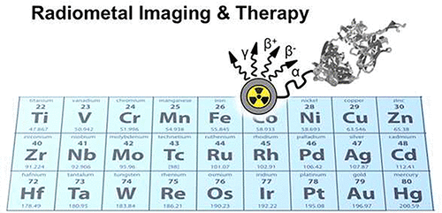
Nuclear medicine is composed of two complementary areas, imaging and therapy. Positron emission tomography (PET) and single-photon imaging, including single-photon emission computed tomography (SPECT), comprise the imaging component of nuclear medicine. These areas are distinct in that they exploit different nuclear decay processes and also different imaging technologies. In PET, images are created from the 511 keV photons produced when the positron emitted by a radionuclide encounters an electron and is annihilated. In contrast, in single-photon imaging, images are created from the γ rays (and occasionally X-rays) directly emitted by the nucleus. Therapeutic nuclear medicine uses particulate radiation such as Auger or conversion electrons or β- or α particles. All three of these technologies are linked by the requirement that the radionuclide must be attached to a suitable vector that can deliver it to its target. It is imperative that the radionuclide remain attached to the vector before it is delivered to its target as well as after it reaches its target or else the resulting image (or therapeutic outcome) will not reflect the biological process of interest. Radiochemistry is at the core of this process, and radiometals offer radiopharmaceutical chemists a tremendous range of options with which to accomplish these goals. They also offer a wide range of options in terms of radionuclide half-lives and emission properties, providing the ability to carefully match the decay properties with the desired outcome. This Review provides an overview of some of the ways this can be accomplished as well as several historical examples of some of the limitations of earlier metalloradiopharmaceuticals and the ways that new technologies, primarily related to radionuclide production, have provided solutions to these problems.
中文翻译:

用于成像和治疗的放射性过渡金属。
核医学由两个互补领域组成,即成像和治疗。正电子发射断层扫描(PET)和单光子成像,包括单光子发射计算机断层摄影(SPECT),构成了核医学的成像组件。这些领域的不同之处在于它们利用不同的核衰变过程以及不同的成像技术。在PET中,图像是由放射性核素发射的正电子遇到电子并被when灭时产生的511 keV光子产生的。相反,在单光子成像中,图像是由原子核直接发射的γ射线(有时是X射线)创建的。治疗性核医学使用诸如俄歇或转换电子或β-或α粒子之类的微粒辐射。所有这三项技术的联系是放射性核素必须附着在合适的载体上,才能将其传递至目标。至关重要的是,放射性核素在被递送至其靶标之前以及到达其靶标之后必须保持附着在载体上,否则所得图像(或治疗结果)将不会反映出所关注的生物学过程。放射性化学是该过程的核心,放射性金属为放射性药物化学家提供了实现这些目标的多种选择。在放射性核素的半衰期和发射特性方面,它们还提供了广泛的选择范围,使人们能够仔细地将衰变特性与所需的结果进行匹配。
更新日期:2018-10-09
中文翻译:
用于成像和治疗的放射性过渡金属。
核医学由两个互补领域组成,即成像和治疗。正电子发射断层扫描(PET)和单光子成像,包括单光子发射计算机断层摄影(SPECT),构成了核医学的成像组件。这些领域的不同之处在于它们利用不同的核衰变过程以及不同的成像技术。在PET中,图像是由放射性核素发射的正电子遇到电子并被when灭时产生的511 keV光子产生的。相反,在单光子成像中,图像是由原子核直接发射的γ射线(有时是X射线)创建的。治疗性核医学使用诸如俄歇或转换电子或β-或α粒子之类的微粒辐射。所有这三项技术的联系是放射性核素必须附着在合适的载体上,才能将其传递至目标。至关重要的是,放射性核素在被递送至其靶标之前以及到达其靶标之后必须保持附着在载体上,否则所得图像(或治疗结果)将不会反映出所关注的生物学过程。放射性化学是该过程的核心,放射性金属为放射性药物化学家提供了实现这些目标的多种选择。在放射性核素的半衰期和发射特性方面,它们还提供了广泛的选择范围,使人们能够仔细地将衰变特性与所需的结果进行匹配。


 京公网安备 11010802027423号