当前位置:
X-MOL 学术
›
ACS Cent. Sci.
›
论文详情
Our official English website, www.x-mol.net, welcomes your
feedback! (Note: you will need to create a separate account there.)
In Vivo Two-Photon Voltage Imaging with Sulfonated Rhodamine Dyes
ACS Central Science ( IF 12.7 ) Pub Date : 2018-10-08 00:00:00 , DOI: 10.1021/acscentsci.8b00422 Rishikesh U. Kulkarni , Matthieu Vandenberghe , Martin Thunemann , Feroz James , Ole A. Andreassen , Srdjan Djurovic , Anna Devor 1 , Evan W. Miller
ACS Central Science ( IF 12.7 ) Pub Date : 2018-10-08 00:00:00 , DOI: 10.1021/acscentsci.8b00422 Rishikesh U. Kulkarni , Matthieu Vandenberghe , Martin Thunemann , Feroz James , Ole A. Andreassen , Srdjan Djurovic , Anna Devor 1 , Evan W. Miller
Affiliation

|
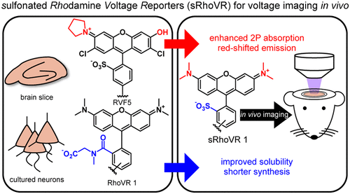
Optical methods that rely on fluorescence for mapping changes in neuronal membrane potential in the brains of awake animals provide a powerful way to interrogate the activity of neurons that underlie neural computations ranging from sensation and perception to learning and memory. To achieve this goal, fluorescent indicators should be bright, highly sensitive to small changes in membrane potential, nontoxic, and excitable with infrared light. We report a new class of fluorescent, voltage-sensitive dyes: sulfonated rhodamine voltage reporters (sRhoVR), synthetic fluorophores with high voltage sensitivity, excellent two-photon performance, and compatibility in intact mouse brains. sRhoVR dyes are based on a tetramethyl rhodamine fluorophore coupled to a phenylenevinylene molecular wire/diethyl aniline voltage-sensitive domain. When applied to cells, sRhoVR dyes localize to the plasma membrane and respond to membrane depolarization with a fluorescence increase. The best of the new dyes, sRhoVR 1, displays a 44% ΔF/F increase in fluorescence per 100 mV change, emits at 570 nm, and possesses excellent two-photon absorption of approximately 200 GM at 840 nm. sRhoVR 1 can detect action potentials in cultured rat hippocampal neurons under both single- and two-photon illumination with sufficient speed and sensitivity to report on action potentials in single trials, without perturbing underlying physiology or membrane properties. The combination of speed, sensitivity, and brightness under two-photon illumination makes sRhoVR 1 a promising candidate for in vivo imaging in intact brains. We show sRhoVR powerfully complements electrode-based modes of neuronal activity recording in the mouse brain by recording neuronal transmembrane potentials from the neuropil of layer 2/3 of the mouse barrel cortex in concert with extracellularly recorded local field potentials (LFPs). sRhoVR imaging reveals robust depolarization in response to whisker stimulation; concurrent electrode recordings reveal negative deflections in the LFP recording, consistent with the canonical thalamocortical response. Importantly, sRhoVR 1 can be applied in mice with chronic optical windows, presaging its utility in dissecting and resolving voltage dynamics using two-photon functional imaging in awake, behaving animals.
中文翻译:

磺化若丹明染料的体内双光子电压成像
依靠荧光来绘制清醒动物大脑中神经元膜电位变化的光学方法,提供了一种强大的方法来询问神经元的活动,这些活动是从感觉和知觉到学习和记忆的神经计算的基础。为实现此目标,荧光指示剂应明亮,对膜电位的微小变化高度敏感,无毒且可被红外光激发。我们报告了一种新型的对电压敏感的荧光染料:磺化罗丹明电压报告分子(sRhoVR),具有高电压敏感性,出色的两光子性能以及在完整小鼠大脑中的相容性的合成荧光团。sRhoVR染料基于偶联至亚苯基亚乙烯基分子线/二乙基苯胺电压敏感域的四甲基若丹明荧光团。当应用于细胞时,sRhoVR染料位于质膜上,并通过荧光增强对膜去极化作出反应。最好的新型染料sRhoVR 1显示出44%的ΔF / F每100 mV变化会增加荧光,在570 nm处发射,并在840 nm处具有约200 GM的出色的双光子吸收。sRhoVR 1可以在单光和双光子照射下以足够的速度和灵敏度检测培养的大鼠海马神经元中的动作电位,以在单次试验中报告动作电位,而不会影响潜在的生理或膜特性。sRhoVR 1在双光子照明下的速度,灵敏度和亮度的结合使sRhoVR 1成为体内有希望的候选者在完整的大脑中成像。我们显示sRhoVR通过记录小鼠桶状皮层的2/3层的神经纤维与细胞外记录的局部场电位(LFPs)的神经元跨膜电位,对小鼠脑中神经元活动记录的基于电极的模式进行了有力补充。sRhoVR成像揭示了对晶须刺激的强烈去极化;并发的电极记录显示出LFP记录中的负偏斜,与典型的丘脑皮质反应一致。重要的是,sRhoVR 1可以应用于具有慢性光学窗口的小鼠,从而预示了它在清醒,表现良好的动物中使用双光子功能成像分析和解决电压动态方面的效用。
更新日期:2018-10-08
中文翻译:

磺化若丹明染料的体内双光子电压成像
依靠荧光来绘制清醒动物大脑中神经元膜电位变化的光学方法,提供了一种强大的方法来询问神经元的活动,这些活动是从感觉和知觉到学习和记忆的神经计算的基础。为实现此目标,荧光指示剂应明亮,对膜电位的微小变化高度敏感,无毒且可被红外光激发。我们报告了一种新型的对电压敏感的荧光染料:磺化罗丹明电压报告分子(sRhoVR),具有高电压敏感性,出色的两光子性能以及在完整小鼠大脑中的相容性的合成荧光团。sRhoVR染料基于偶联至亚苯基亚乙烯基分子线/二乙基苯胺电压敏感域的四甲基若丹明荧光团。当应用于细胞时,sRhoVR染料位于质膜上,并通过荧光增强对膜去极化作出反应。最好的新型染料sRhoVR 1显示出44%的ΔF / F每100 mV变化会增加荧光,在570 nm处发射,并在840 nm处具有约200 GM的出色的双光子吸收。sRhoVR 1可以在单光和双光子照射下以足够的速度和灵敏度检测培养的大鼠海马神经元中的动作电位,以在单次试验中报告动作电位,而不会影响潜在的生理或膜特性。sRhoVR 1在双光子照明下的速度,灵敏度和亮度的结合使sRhoVR 1成为体内有希望的候选者在完整的大脑中成像。我们显示sRhoVR通过记录小鼠桶状皮层的2/3层的神经纤维与细胞外记录的局部场电位(LFPs)的神经元跨膜电位,对小鼠脑中神经元活动记录的基于电极的模式进行了有力补充。sRhoVR成像揭示了对晶须刺激的强烈去极化;并发的电极记录显示出LFP记录中的负偏斜,与典型的丘脑皮质反应一致。重要的是,sRhoVR 1可以应用于具有慢性光学窗口的小鼠,从而预示了它在清醒,表现良好的动物中使用双光子功能成像分析和解决电压动态方面的效用。


 京公网安备 11010802027423号