Our official English website, www.x-mol.net, welcomes your feedback! (Note: you will need to create a separate account there.)
Enhancement of optical resolution in three-dimensional refractive-index tomograms of biological samples by employing micromirror-embedded coverslips†
Lab on a Chip ( IF 6.1 ) Pub Date : 2018-10-04 00:00:00 , DOI: 10.1039/c8lc00880a Seungwoo Shin 1, 2, 3, 4, 5 , Jihye Kim 3, 4, 6, 7 , Je-Ryung Lee 8, 9, 10, 11 , Eun-chae Jeon 11, 12, 13, 14 , Tae-Jin Je 8, 9, 10, 11 , Wonhee Lee 1, 2, 3, 4, 6 , YongKeun Park 1, 2, 3, 4, 5
Lab on a Chip ( IF 6.1 ) Pub Date : 2018-10-04 00:00:00 , DOI: 10.1039/c8lc00880a Seungwoo Shin 1, 2, 3, 4, 5 , Jihye Kim 3, 4, 6, 7 , Je-Ryung Lee 8, 9, 10, 11 , Eun-chae Jeon 11, 12, 13, 14 , Tae-Jin Je 8, 9, 10, 11 , Wonhee Lee 1, 2, 3, 4, 6 , YongKeun Park 1, 2, 3, 4, 5
Affiliation

|
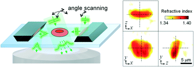
Optical diffraction tomography (ODT) enables the reconstruction of the three-dimensional (3D) refractive-index (RI) distribution of a biological cell, which provides invaluable information for cellular and subcellular structures in a non-invasive manner. However, ODT suffers from an inferior axial resolution, due to the limited accessible angles imposed by the numerical aperture of the objective lens. In this study, we propose and experimentally demonstrate an approach to enhance the 3D reconstruction performance in ODT. By employing trapezoidal micromirrors, side scattered signals from the sample are measured for various side plane-wave-illumination angles. By combining the side scattered fields with the forward scattered fields, the axial resolution and 3D image quality of ODT are improved, without changing optical instruments. The feasibility and applicability of the proposed method are demonstrated by reconstructing the 3D RI distribution of a red blood cell and HeLa cells in hydrogel. We also present systematic analyses of the improved 3D imaging performance using numerical simulations and experimental measurements for the 3D transfer function, a point object, and a microsphere. The analyses demonstrate an improved axial resolution of 0.31 μm, 4.8 times smaller than that of the conventional method. The proposed method enables the non-invasive and accurate 3D imaging of 3D cultured cells, which is crucial for cell biology studies.
中文翻译:

通过使用微镜嵌入式盖玻片来增强生物样品的三维折射率X线断层图中的光学分辨率†
光学衍射断层扫描(ODT)能够重建生物细胞的三维(3D)折射率(RI)分布,从而以无创方式为细胞和亚细胞结构提供了宝贵的信息。然而,由于物镜的数值孔径所施加的有限的可到达角度,ODT的轴向分辨率较低。在这项研究中,我们提出并通过实验演示了一种增强ODT中3D重建性能的方法。通过使用梯形微镜,针对各种侧平面波照明角度,测量了来自样品的侧向散射信号。通过将侧面散射场和前向散射场结合在一起,ODT的轴向分辨率和3D图像质量得以提高,而无需更换光学仪器。通过重构水凝胶中红细胞和HeLa细胞的3D RI分布证明了该方法的可行性和适用性。我们还使用数值模拟和3D传递函数,点对象和微球体的实验测量结果,对改进的3D成像性能进行了系统分析。分析表明,轴向分辨率提高了0.31μm,是传统方法的4.8倍。所提出的方法能够对3D培养的细胞进行无创且准确的3D成像,这对于细胞生物学研究至关重要。我们还使用数值模拟和3D传递函数,点对象和微球体的实验测量结果,对改进的3D成像性能进行了系统分析。分析显示轴向分辨率提高了0.31μm,比传统方法的轴向分辨率小4.8倍。所提出的方法能够对3D培养的细胞进行无创且准确的3D成像,这对于细胞生物学研究至关重要。我们还使用数值模拟和3D传递函数,点对象和微球体的实验测量结果,对改进的3D成像性能进行了系统分析。分析显示轴向分辨率提高了0.31μm,比传统方法的轴向分辨率小4.8倍。所提出的方法能够对3D培养的细胞进行无创且准确的3D成像,这对于细胞生物学研究至关重要。
更新日期:2018-10-04
中文翻译:

通过使用微镜嵌入式盖玻片来增强生物样品的三维折射率X线断层图中的光学分辨率†
光学衍射断层扫描(ODT)能够重建生物细胞的三维(3D)折射率(RI)分布,从而以无创方式为细胞和亚细胞结构提供了宝贵的信息。然而,由于物镜的数值孔径所施加的有限的可到达角度,ODT的轴向分辨率较低。在这项研究中,我们提出并通过实验演示了一种增强ODT中3D重建性能的方法。通过使用梯形微镜,针对各种侧平面波照明角度,测量了来自样品的侧向散射信号。通过将侧面散射场和前向散射场结合在一起,ODT的轴向分辨率和3D图像质量得以提高,而无需更换光学仪器。通过重构水凝胶中红细胞和HeLa细胞的3D RI分布证明了该方法的可行性和适用性。我们还使用数值模拟和3D传递函数,点对象和微球体的实验测量结果,对改进的3D成像性能进行了系统分析。分析表明,轴向分辨率提高了0.31μm,是传统方法的4.8倍。所提出的方法能够对3D培养的细胞进行无创且准确的3D成像,这对于细胞生物学研究至关重要。我们还使用数值模拟和3D传递函数,点对象和微球体的实验测量结果,对改进的3D成像性能进行了系统分析。分析显示轴向分辨率提高了0.31μm,比传统方法的轴向分辨率小4.8倍。所提出的方法能够对3D培养的细胞进行无创且准确的3D成像,这对于细胞生物学研究至关重要。我们还使用数值模拟和3D传递函数,点对象和微球体的实验测量结果,对改进的3D成像性能进行了系统分析。分析显示轴向分辨率提高了0.31μm,比传统方法的轴向分辨率小4.8倍。所提出的方法能够对3D培养的细胞进行无创且准确的3D成像,这对于细胞生物学研究至关重要。


 京公网安备 11010802027423号