PLOS ONE ( IF 2.9 ) Pub Date : 2018-09-19 , DOI: 10.1371/journal.pone.0203940 Ye Ra Choi , Jin Wook Chung , Mi Hye Yu , Myungsu Lee , Jung Hoon Kim
|
Objective
To evaluate the accuracy of CT for small, hypervascular hepatocellular carcinomas (HCCs) and assess the enhancement patterns on CT.
Materials and methods
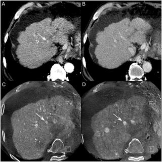
Ninety-nine patients who underwent cone-beam CT hepatic arteriography (CBCT-HA) during initial chemoembolization for HCC suspected on CT were enrolled in this study. A total of 297 hypervascular HCCs (142 ≥ 1 cm, 155 < 1 cm) were confirmed as HCCs based on two-year follow-up CT and CBCT-HA images. During the two-year follow-up, pre-existing hypervascular foci on CBCT-HA were regarded as HCCs at the initial presentation. Two radiologists categorized HCCs according to the following enhancement patterns on CT: type I, arterial enhancement and washout; type II, arterial enhancement without washout; and type III, no arterial enhancement. Two blinded reviewers rated the possibility of HCC.
Results
For the 297 HCCs, the enhancement patterns according to size were as follows: type I ≥1 cm in 114 HCCs; type I <1 cm in 40 HCCs; type II ≥1 cm in 16 HCCs; type II <1 cm in 37 HCCs; type III ≥1 cm in 12 HCCs; and type III <1 cm in 10 HCCs. The remaining 68 HCCs (22.9%) were not detected on CT. The detection rates of HCCs ≥ 1 cm were 83.1%, 76.8%, and 83.1% in the formal report for reviewer 1 and reviewer 2. In comparison, the detection rates of HCCs < 1 cm were 20.6%, 17.4%, and 17.4% in the formal report for reviewer 1 and reviewer 2.
Conclusion
Many subcentimeter sized hypervascular HCCs were frequently missed or not evident on CT at the initial diagnostic workup. CT has limitations for diagnosing HCCs that are <1 cm in size or have atypical enhancement patterns.
中文翻译:
增强动态CT对小型高血管性肝细胞癌的诊断准确性和动态增强模式的评估:锥束CT肝动脉造影两年随访的结果
客观的
评估CT对小型高血管性肝细胞癌(HCC)的准确性,并评估CT的增强模式。
材料和方法
本研究招募了99例在最初的肝癌化疗栓塞期间接受锥束CT肝动脉造影(CBCT-HA)的患者。根据为期两年的CT和CBCT-HA随访图像,共确认297例高血管HCC(142≥1 cm,155 <1 cm)为HCC。在为期两年的随访中,最初出现时,CBCT-HA上已存在的高血管灶被视为肝癌。两名放射科医师根据以下CT上的增强模式对HCC进行了分类:I型,动脉增强和冲洗;II型,无冲洗的动脉增强;和III型,无动脉增强。两名不知情的评论者对HCC的可能性进行了评估。
结果
对于297个HCC,根据大小的增强模式如下:114个HCC中的I型≥1cm; I型≥1cm。在40个HCC中I型<1厘米; II型≥1cm在16个HCC中; II型<1厘米,在37个HCC中; 在12个HCC中III型≥1厘米; 并在10个HCC中键入III <1 cm。在CT上未检测到其余68例HCC(22.9%)。在审稿人1和审稿人2的正式报告中,≥1 cm的HCC的检出率分别为83.1%,76.8%和83.1%。相比之下,<1 cm的HCC的检出率分别为20.6%,17.4%和17.4%。在针对审稿人1和审稿人2的正式报告中。
结论
曼ÿ亚厘米级尺寸的富血肝癌被经常错过的或不按CT明显在初始诊断检查。CT在诊断尺寸小于1厘米或具有非典型增强型肝癌时有局限性。


 京公网安备 11010802027423号