Acta Biomaterialia ( IF 9.7 ) Pub Date : 2018-09-16 , DOI: 10.1016/j.actbio.2018.09.020 Harmanvir Ghuman 1 , Carrinton Mauney 2 , Julia Donnelly 3 , Andre R Massensini 4 , Stephen F Badylak 5 , Michel Modo 6
|
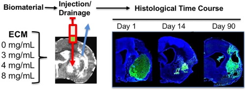
The brain is considered to have a limited capacity to repair damaged tissue and no regenerative capacity following injury. Tissue lost after a stroke is therefore not spontaneously replaced. Extracellular matrix (ECM)-based hydrogels implanted into the stroke cavity can attract endogenous cells. These hydrogels can be formulated at different protein concentrations that govern their rheological and inductive properties. We evaluated histologically 0, 3, 4 and 8 mg/mL of porcine-derived urinary bladder matrix (UBM)-ECM hydrogel concentrations implanted in a 14-day old stroke cavity. Less concentrated hydrogels (3 and 4 mg/mL) were efficiently degraded with a 95% decrease in volume by 90 days, whereas only 32% of the more concentrated and stiffer hydrogel (8 mg/mL) was resorbed. Macrophage infiltration and density within the bioscaffold progressively increased in the less concentrated hydrogels and decreased in the 8 mg/mL hydrogels. The less concentrated hydrogels showed a robust invasion of endothelial cells with neovascularization. No neovascularization occurred with the stiffer hydrogel. Invasion of neural cells increased with time in all hydrogel concentrations. Differentiation of neural progenitors into mature neurons with axonal projections was evident, as well as a robust invasion of oligodendrocytes. However, relatively few astrocytes were present in the ECM hydrogel, although some were present in the newly forming tissue between degrading scaffold patches. Implantation of an ECM hydrogel partially induced neural tissue restoration, but a more complete understanding is required to evaluate its potential therapeutic application.
Statement of Significance
Extracellular matrix hydrogel promotes tissue regeneration in many peripheral soft tissues. However, the brain has generally been considered to lack the potential for tissue regeneration. We here demonstrate that tissue regeneration in the brain can be achieved using implantation of ECM hydrogel into a tissue cavity. We here demonstrate that a structure-function relationship is key to promote tissue regeneration in the brain. Specifically, weaker hydrogels that were retained in the cavity underwent an efficient biodegradation within 14 days post-implantation to promote a tissue restoration within the lesion cavity. In contrast, stiffer ECM hydrogel only underwent minor biodegradation and did not lead to a tissue restoration. Inductive hydrogels weaker than brain tissue provide the appropriate condition to promote an endogenous regenerative response that restores tissue in a cavity. This approach offers new avenues for the future treatment of chronic tissue damage caused by stroke and other acute brain injuries.
中文翻译:
ECM 水凝胶的生物降解促进中风大鼠模型的内源性脑组织恢复
大脑被认为修复受损组织的能力有限,并且在受伤后没有再生能力。因此,中风后丢失的组织不会自发替换。植入中风腔内的基于细胞外基质(ECM)的水凝胶可以吸引内源性细胞。这些水凝胶可以配制为不同的蛋白质浓度,以控制其流变学和诱导特性。我们对植入 14 天的中风空腔中的 0、3、4 和 8 mg/mL 猪源性膀胱基质 (UBM)-ECM 水凝胶浓度进行了组织学评估。浓度较低的水凝胶(3 和 4 mg/mL)可有效降解,90 天体积减少 95%,而浓度较高且较硬的水凝胶(8 mg/mL)仅 32% 被再吸收。生物支架内的巨噬细胞浸润和密度在浓度较低的水凝胶中逐渐增加,在 8 mg/mL 水凝胶中逐渐减少。浓度较低的水凝胶显示出内皮细胞的强烈侵袭和新血管形成。较硬的水凝胶没有发生新血管形成。在所有水凝胶浓度下,神经细胞的侵袭随着时间的推移而增加。神经祖细胞明显分化为具有轴突投射的成熟神经元,并且少突胶质细胞的强烈侵袭。然而,ECM 水凝胶中存在的星形胶质细胞相对较少,尽管一些星形胶质细胞存在于降解支架补片之间新形成的组织中。ECM 水凝胶的植入部分诱导神经组织恢复,但需要更全面的理解来评估其潜在的治疗应用。
重要性声明
细胞外基质水凝胶促进许多外周软组织的组织再生。然而,人们普遍认为大脑缺乏组织再生的潜力。我们在此证明,通过将 ECM 水凝胶植入组织腔中可以实现大脑中的组织再生。我们在这里证明结构-功能关系是促进大脑组织再生的关键。具体来说,保留在腔内的较弱的水凝胶在植入后 14 天内进行有效的生物降解,以促进病变腔内的组织恢复。相比之下,较硬的 ECM 水凝胶仅经历轻微的生物降解,并且不会导致组织修复。比脑组织弱的感应水凝胶提供了适当的条件来促进内源性再生反应,从而恢复腔内的组织。这种方法为未来治疗中风和其他急性脑损伤引起的慢性组织损伤提供了新途径。


 京公网安备 11010802027423号