Acta Biomaterialia ( IF 9.7 ) Pub Date : 2018-09-13 , DOI: 10.1016/j.actbio.2018.09.010 Rachel M. Cahalane , Hilary E. Barrett , Julie M. O'Brien , Eamon G. Kavanagh , Michael A. Moloney , Michael T. Walsh
|
Calcification is associated with decreased atherosclerotic plaque stability and increased failures rates for endovascular interventions. Computational efforts have sought to elucidate the relationship between calcification and plaque rupture in addition to predicting tissue response during aggressive revascularisation techniques. However, calcified material properties are currently estimated and may not reflect real tissue conditions. The objective of this study is to correlate calcification mechanical properties with three radiographic density groups obtained from corresponding Computed Tomography (CT) images.
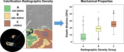
Seventeen human plaques extracted from carotid (n=10) and peripheral lower limb (n=7) arteries were examined using micro-computed tomography (µCT), simultaneously locating the calcified deposits within their internal structure and quantifying their densities. Three radiographic density groups were defined based on the sample density distribution: (A) 130-299.99 Hounsfield Units (HU), (B) 300-449.99HU and (C) >450HU. Nanoindentation was employed to determine the Elastic Modulus (E) and Hardness (H) values within the three density groups.
Results reveal a clear distinction between mechanical properties with respect to radiographic density groups (p<0.0005). No significant differences exist in the density-specific behaviours observed between carotid and peripheral samples. Previously defined calcification classifications indicate an association with specific radiographic density patterns. Scanning Electron Microscopy (SEM) examination revealed that density group A regions consist of both calcified and non-calcified tissues. Further research is required to define the radiographic thresholds which identify varying degrees of tissue calcification.
This study demonstrates that the mechanical properties of fully mineralised atherosclerotic calcification emulate that of bone tissues (17-25GPa), affording computational models with accurate material parameters.
Statement of Significance
Global mechanical characterisation techniques disregard the heterogeneous nature of atherosclerotic lesions. Previous nanoindentation results for carotid calcifications have displayed a wide range. This study evaluates calcification properties with respect to radiographic density obtained from Micro-CT images. This is the first work to characterise calcifications from peripheral lower limb arteries using nanoindentation. Results demonstrate a strong positive correlation between radiographic density and calcification mechanical properties. Characterising calcifications using their density values provides clarity on the variation in published properties for calcified tissues. Furthermore, this study confirms the hypothesis that fully calcified plaque tissue behaviours similar to that of bone. Appropriate material parameters for calcified tissues can now be employed in computational simulations.
中文翻译:
动脉粥样硬化钙化的力学性能与射线密度的关系:纳米压痕方法。
钙化与动脉粥样硬化斑块稳定性降低和血管内干预失败率增加相关。除了预测积极的血管重建技术期间的组织反应以外,计算工作还试图阐明钙化和斑块破裂之间的关系。但是,目前估计的是钙化的材料特性,可能无法反映真实的组织状况。这项研究的目的是将钙化力学性能与从相应的计算机断层扫描(CT)图像获得的三个射线照相密度组相关联。
使用微型计算机断层扫描(µCT)检查了从颈动脉(n = 10)和周围下肢动脉(n = 7)动脉中提取的17个人类斑块,同时在其内部结构中定位了钙化沉积物并量化了其密度。根据样品密度分布定义了三个射线照相密度组:(A)130-299.99豪恩斯单位(HU),(B)300-449.99HU和(C)> 450HU。纳米压痕用于确定三个密度组内的弹性模量(E)和硬度(H)值。
结果表明,相对于射线照相密度组,机械性能之间存在明显区别(p <0.0005)。在颈动脉和外周血样品之间观察到的密度特异性行为方面没有显着差异。先前定义的钙化分类表明与特定的射线照相密度模式相关。扫描电子显微镜(SEM)检查显示,密度A组区域由钙化组织和非钙化组织组成。需要进一步的研究来确定可识别组织钙化程度不同的放射线阈值。
这项研究表明,完全矿化的动脉粥样硬化钙化的机械性能可以模拟骨骼组织(17-25GPa)的力学性能,从而提供具有精确材料参数的计算模型。
重要声明
全球机械表征技术忽略了动脉粥样硬化病变的异质性。先前关于颈动脉钙化的纳米压痕结果显示了广泛的范围。这项研究评估了有关从Micro-CT图像获得的射线照相密度的钙化特性。这是使用纳米压痕技术表征下肢周围动脉钙化的第一项工作。结果表明射线照相密度和钙化机械性能之间有很强的正相关性。使用钙离子密度值表征钙化,可以清楚地显示钙化组织的已公布属性的变化。此外,这项研究证实了完全钙化斑块组织行为与骨骼相似的假说。


 京公网安备 11010802027423号