Nature Methods ( IF 36.1 ) Pub Date : 2018-09-10 , DOI: 10.1038/s41592-018-0115-y Tianyu Wang 1 , Dimitre G Ouzounov 1 , Chunyan Wu 1 , Nicholas G Horton 1 , Bin Zhang 2 , Cheng-Hsun Wu 2 , Yanping Zhang 2, 3 , Mark J Schnitzer 2, 3 , Chris Xu 1
|
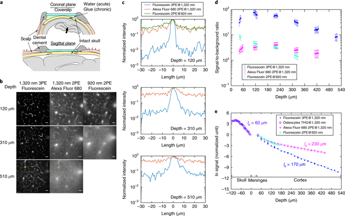
Optical imaging through the intact mouse skull is challenging because of skull-induced aberrations and scattering. We found that three-photon excitation provided improved optical sectioning compared with that obtained with two-photon excitation, even when we used the same excitation wavelength and imaging system. Here we demonstrate three-photon imaging of vasculature through the adult mouse skull at >500-μm depth, as well as GCaMP6s calcium imaging over weeks in cortical layers 2/3 and 4 in awake mice, with 8.5 frames per second and a field of view spanning hundreds of micrometers.
中文翻译:
通过完整头骨对小鼠大脑结构和功能进行三光子成像
由于头骨引起的像差和散射,通过完整的小鼠头骨进行光学成像具有挑战性。我们发现,即使我们使用相同的激发波长和成像系统,与双光子激发获得的光学切片相比,三光子激发提供了改进的光学切片。在这里,我们展示了通过成年小鼠头骨在 >500-μm 深度处进行脉管系统的三光子成像,以及在清醒小鼠的皮质层 2/3 和 4 中数周内进行的 GCaMP6s 钙成像,每秒 8.5 帧,视野为视野跨越数百微米。


 京公网安备 11010802027423号