Cellular Signalling ( IF 4.4 ) Pub Date : 2018-09-01 , DOI: 10.1016/j.cellsig.2018.08.021 Ela Markovsky 1 , Elisa de Stanchina 2 , Aryeh Itzkowitz 3 , Adriana Haimovitz-Friedman 1 , Susan A Rotenberg 4
|
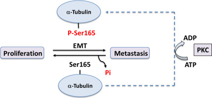
Engineered overexpression of protein kinase Cα (PKCα) is known to phosphorylate Ser165 in α-tubulin resulting in stimulated microtubule dynamics and cell motility, and activation of an epithelial-mesenchymal transition (EMT) in non-transformed human breast cells. Here it is shown that endogenous phosphorylation of native α-tubulin in two metastatic breast cell lines, MDA-MB-231-LM2–4175 and MDA-MB-468 is detected at PKC phosphorylation sites. α-Tubulin mutants that simulated phosphorylated (S165D) or non-phosphorylated (S165 N) states were stably expressed in MDA-MB-231-LM2–4175 cells. The S165D-α-tubulin mutant engendered expression of the EMT biomarker N-cadherin, whereas S165 N-α-tubulin suppressed N-cadherin and induced E-cadherin expression, revealing a ‘cadherin switch’. S165 N-α-tubulin engendered more rapid passage through the cell cycle, induced shorter spindle fibers and exhibited more rapid proliferation. In nude mice injected with MDA-MB-231-LM2–4175 cells, cells expressing S165 N-α-tubulin (but not the S165D mutant) produced hyper-proliferative lung tumors with increased tumor incidence and higher Ki67 expression. These results implicate the phosphorylation state of Ser165 in α-tubulin as a PKC-regulated molecular switch that causes breast cells to exhibit either EMT characteristics or hyper-proliferation. Evaluation of genomic databases of human tumors strengthens the clinical significance of these findings.
中文翻译:
α-微管蛋白中 Ser165 的磷酸化状态是控制人类乳腺肿瘤增殖的切换开关
已知蛋白激酶 Cα (PKCα) 的工程过度表达可磷酸化α-微管蛋白中的Ser 165 ,从而刺激微管动力学和细胞运动,并激活未转化的人乳腺细胞中的上皮间质转化 (EMT)。此处显示,在两种转移性乳腺细胞系 MDA-MB-231-LM2-4175 和 MDA-MB-468 中,在 PKC 磷酸化位点检测到天然 α-微管蛋白的内源磷酸化。模拟磷酸化 (S165D) 或非磷酸化 (S165 N) 状态的 α-微管蛋白突变体在 MDA-MB-231-LM2–4175 细胞中稳定表达。S165D-α-微管蛋白突变体引起EMT生物标志物N-钙粘蛋白的表达,而S165 N-α-微管蛋白抑制N-钙粘蛋白并诱导E-钙粘蛋白表达,揭示了“钙粘蛋白开关”。S165 N-α-微管蛋白导致更快速地通过细胞周期,诱导更短的纺锤体纤维并表现出更快速的增殖。在注射 MDA-MB-231-LM2-4175 细胞的裸鼠中,表达 S165 N-α-微管蛋白(但不是 S165D 突变体)的细胞产生过度增殖性肺肿瘤,肿瘤发生率增加,Ki67 表达更高。这些结果暗示α-微管蛋白中Ser 165的磷酸化状态作为 PKC 调节的分子开关,导致乳腺细胞表现出 EMT 特征或过度增殖。对人类肿瘤基因组数据库的评估增强了这些发现的临床意义。


 京公网安备 11010802027423号