Acta Biomaterialia ( IF 9.4 ) Pub Date : 2018-08-31 , DOI: 10.1016/j.actbio.2018.08.039 Jevin G. Meyerink , Divya Kota , Scott T. Wood , Grant A. Crawford
|
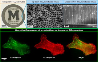
The therapeutic applications of titanium dioxide nanotubes as osteogenic surface treatments for titanium-based implants are largely due to the finely tunable physical characteristics of these nanostructures. As these characteristics change, so does the cellular response, yet the exact mechanisms for this relationship remains largely undefined. We present a novel TiO2 NT imaging platform that is suitable for use with live-cell imaging techniques, thereby enabling, for the first time, dynamic investigation of those mechanisms. In this work, fabrication methods for producing transparent TiO2 NTs with diameters of 56 ± 6 nm, 75 ± 7 nm, 92 ± 9 nm, and 116 ± 10 nm are described. To demonstrate the diagnostic potential of these TiO2 NT imaging platforms, the focal adhesion protein vinculin and actin cytoskeletal filaments were fluorescently tagged in osteoblasts and real-time, high-resolution fluorescent microscopy of live-cell interactions with TiO2 NT substrates were observed. The scope of such a platform is expected to extend far beyond the current proof-of-concept, with great potential for addressing the dynamic response of cells interacting with nanostructured substrates.
Statement of Significance
Titanium dioxide (TiO2) nanotubes are known to strongly enhance bone/mesenchymal stem cell behavior and, consequently, have gained attention as potential osteogenic surface treatments for titanium-bone implants. The exact mechanism by which TiO2 nanotubes influence cellular function remains controversial, partly due to limitations in existing cellular imaging methods with opaque substrates. This work identifies fabrication conditions for the successful production of transparent TiO2 nanotube arrays with tailorable diameters, as well as their functionality with pre-osteoblast mouse cells (MC3T3-E1) transfected with fluorescent focal adhesion protein vinculin and cytoskeletal filament actin. We demonstrate a means of recording live-cell, cell-substrate interaction mechanisms via high-resolution fluorescent microscopy and customizable, transparent TiO2 nanotubes to begin defining the relationship between TiO2 nanotube features and cell function.
中文翻译:
透明二氧化钛纳米管:加工,表征,并在建立细胞反应机制中的应用。
二氧化钛纳米管作为钛基植入物的成骨表面处理的治疗应用很大程度上归因于这些纳米结构的精细可调的物理特性。随着这些特性的改变,细胞反应也随之改变,但是这种关系的确切机制在很大程度上仍然不确定。我们提出了一种新颖的TiO 2 NT成像平台,适用于活细胞成像技术,从而首次实现了对这些机制的动态研究。在这项工作中,描述了制备直径为56±6 nm,75±7 nm,92±9 nm和116±10 nm的透明TiO 2 NTs的制造方法。证明这些TiO 2的诊断潜力NT成像平台,黏着斑蛋白纽蛋白和肌动蛋白细胞骨架丝在成骨细胞中进行了荧光标记,并实时,高分辨率荧光显微镜观察了活细胞与TiO 2 NT底物的相互作用。预计该平台的范围将远远超出当前的概念验证,具有解决与纳米结构化基材相互作用的细胞动态响应的巨大潜力。
重要声明
众所周知,二氧化钛(TiO 2)纳米管可显着增强骨骼/间充质干细胞的行为,因此,作为钛骨植入物的潜在成骨表面处理剂已引起人们的关注。TiO 2纳米管影响细胞功能的确切机制仍存在争议,部分原因是现有的不透明基材细胞成像方法的局限性。这项工作确定了成功生产透明TiO 2的制造条件具有可定制直径的纳米管阵列,以及它们在用荧光粘着斑粘附蛋白纽蛋白和细胞骨架细丝肌动蛋白转染的成骨前小鼠细胞(MC3T3-E1)中的功能。我们展示了一种通过高分辨率荧光显微镜和可定制的透明TiO 2纳米管记录活细胞,细胞-基质相互作用机制的方法,从而开始定义TiO 2纳米管特征与细胞功能之间的关系。


 京公网安备 11010802027423号