Polymer Degradation and Stability ( IF 5.9 ) Pub Date : 2018-08-15 , DOI: 10.1016/j.polymdegradstab.2018.08.001 Yu-I Hsu , Tetsuji Yamaoka

|
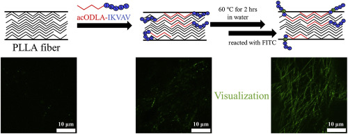
Our ongoing work concerns the modification of poly(L-lactic acid) (PLLA) scaffolds with oligo(lactic acid)-oligo peptide heteroconjugates as a means to improve their bioactivity. Regrettably, the initial cell attachment exhibited by these scaffolds was unsatisfactory, most likely owing to an insufficient density of peptide molecules bound to the outermost surface of the scaffolds. However, to distinguish surface-bound peptides from those embedded in the matrices is difficult by conventional surface analysis techniques such as energy dispersive X-ray spectroscopy and X-ray photoelectron spectroscopy because they detect signals from the bulk surface to a given depth. In the present study, we succeeded in visualizing and quantifying the peptide molecules at the outmost surface only by staining them with an aqueous solution of fluorescein isothiocyanate (FITC). FITC reacts only with the peptides at the surface but not those embedded in the matrices. The uniformity of the peptide coverage can be observed using fluorescence microscopy, and the peptide surface density can be quantified by dissolving the matrices and measuring the intensity of the resultant solution. In addition, by using this quantification method, an effective strategy to increase the peptide density at the PLLA surface was developed. This strategy involves immersing an electrospun nanofibrous sheet composed of PLLA and oligo(D-lactic acid)-IleLysValAlaVal (IKVAV) heteroconjugate (99:1 in weight) in water, warming it to above the glass transition temperature of PLLA (60 °C), and slowly cooling it to room temperature. The surface IKVAV density was highest when treated at 60 °C. During the electrospinning, the IKVAV peptide is mainly embedded in the matrices, but it is exposed to the aqueous phase at the scaffold surface and fixed there by the in-water heating/cooling process.
中文翻译:

可视化和量化固定在基于PLLA的生物材料最外层的生物活性分子
我们正在进行的工作涉及用低聚(乳酸)-低聚肽杂合物修饰聚(L-乳酸)(PLLA)支架,以改善其生物活性。令人遗憾的是,这些支架表现出的初始细胞附着不能令人满意,很可能是由于与支架最外层表面结合的肽分子密度不足所致。但是,通过常规表面分析技术(例如能量色散X射线光谱法和X射线光电子能谱法)很难将表面结合的肽与基质中嵌入的肽区分开来,因为它们会检测从体表到给定深度的信号。在目前的研究中,我们仅通过用异硫氰酸荧光素(FITC)水溶液对肽分子进行染色就可以成功地可视化和量化肽分子的最外层表面。FITC仅与表面上的肽发生反应,而与嵌入基质中的那些不发生反应。可以使用荧光显微镜观察肽覆盖率的均匀性,并且可以通过溶解基质并测量所得溶液的强度来定量肽表面密度。此外,通过使用这种定量方法,开发了一种有效的策略来增加PLLA表面的肽密度。此策略涉及将由PLLA和oligo(D-乳酸)-IleLysValAlaVal(IKVAV)杂合物(重量为99:1)组成的电纺纳米纤维片浸入水中,将其加热至PLLA的玻璃化转变温度(60°C)以上,然后缓慢冷却至室温。当在60°C下处理时,表面IKVAV密度最高。在静电纺丝过程中,IKVAV肽主要嵌入基质中,但在支架表面暴露于水相并通过水中加热/冷却过程固定在那里。


 京公网安备 11010802027423号