Our official English website, www.x-mol.net, welcomes your
feedback! (Note: you will need to create a separate account there.)
Resolution improvement in STED super-resolution microscopy at low power using a phasor plot approach†
Nanoscale ( IF 5.8 ) Pub Date : 2018-08-08 00:00:00 , DOI: 10.1039/c8nr03584a Luwei Wang 1, 2, 3, 4, 5 , Bingling Chen 1, 2, 3, 4, 5 , Wei Yan 1, 2, 3, 4, 5 , Zhigang Yang 1, 2, 3, 4, 5 , Xiao Peng 1, 2, 3, 4, 5 , Danying Lin 1, 2, 3, 4, 5 , Xiaoyu Weng 1, 2, 3, 4, 5 , Tong Ye 6, 7, 8, 9 , Junle Qu 1, 2, 3, 4, 5
Nanoscale ( IF 5.8 ) Pub Date : 2018-08-08 00:00:00 , DOI: 10.1039/c8nr03584a Luwei Wang 1, 2, 3, 4, 5 , Bingling Chen 1, 2, 3, 4, 5 , Wei Yan 1, 2, 3, 4, 5 , Zhigang Yang 1, 2, 3, 4, 5 , Xiao Peng 1, 2, 3, 4, 5 , Danying Lin 1, 2, 3, 4, 5 , Xiaoyu Weng 1, 2, 3, 4, 5 , Tong Ye 6, 7, 8, 9 , Junle Qu 1, 2, 3, 4, 5
Affiliation

|
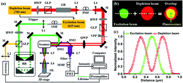
Stimulated emission depletion (STED) microscopy is a powerful super-resolution microscopy technique that has achieved significant results in breaking the resolution limit and relevant applications. In principle, STED super resolution is obtained by stimulated emission partially inhibiting the spontaneous emission in the periphery of a diffraction-limited area. However, very high depletion laser power is generally necessary for the enhancement of imaging resolution, which is harmful to live biological specimens due to its high phototoxicity and photo-bleaching effects. Therefore, further improving the STED resolution at a lower depletion power level has recently attracted increasing interest from researchers in various fields. In this work, a phasor plot approach combined with fluorescence lifetime imaging microscopy (FLIM) is used to resolve the abovementioned problem based on a long- and short-lifetime criterion. Firstly, the time-resolved data obtained by STED-FLIM is converted to the frequency domain via a phasor approach. Next, partial data is extracted according to the information on the phase and amplitude for resolution improvement. Then, fluorescent microspheres (100 nm in diameter) are observed under different depletion powers, resulting in a series of improved resolution through phasor plots. Finally, this method is applied to image human Nup153 in fixed HeLa cells, providing a 86 nm higher resolution than that in traditional STED imaging at a depletion power of 20 mW.
中文翻译:

使用相量图方法在低功率下提高STED超分辨率显微镜的分辨率†
激发发射耗尽(STED)显微镜是一种功能强大的超分辨率显微镜技术,在突破分辨率极限和相关应用方面取得了显著成果。原则上,通过激发发射部分抑制衍射极限区域外围的自发发射来获得STED超分辨率。然而,通常需要非常高的耗尽激光功率来提高成像分辨率,由于其高的光毒性和光漂白作用,这对活生物标本有害。因此,在更低的耗尽功率水平下进一步提高STED分辨率近来引起了各个领域研究人员的越来越多的兴趣。在这项工作中,相量图方法与荧光寿命成像显微镜(FLIM)相结合可用于基于长寿命和短寿命标准来解决上述问题。首先,将STED-FLIM获得的时间分辨数据转换为频域通过相量方法。接下来,根据有关相位和幅度的信息提取部分数据,以提高分辨率。然后,在不同的耗尽功率下观察到荧光微球(直径为100 nm),从而通过相量图获得了一系列改进的分辨率。最后,该方法应用于固定HeLa细胞中的人Nup153成像,在20 mW的耗尽功率下,其分辨率比传统STED成像高86 nm。
更新日期:2018-08-08
中文翻译:

使用相量图方法在低功率下提高STED超分辨率显微镜的分辨率†
激发发射耗尽(STED)显微镜是一种功能强大的超分辨率显微镜技术,在突破分辨率极限和相关应用方面取得了显著成果。原则上,通过激发发射部分抑制衍射极限区域外围的自发发射来获得STED超分辨率。然而,通常需要非常高的耗尽激光功率来提高成像分辨率,由于其高的光毒性和光漂白作用,这对活生物标本有害。因此,在更低的耗尽功率水平下进一步提高STED分辨率近来引起了各个领域研究人员的越来越多的兴趣。在这项工作中,相量图方法与荧光寿命成像显微镜(FLIM)相结合可用于基于长寿命和短寿命标准来解决上述问题。首先,将STED-FLIM获得的时间分辨数据转换为频域通过相量方法。接下来,根据有关相位和幅度的信息提取部分数据,以提高分辨率。然后,在不同的耗尽功率下观察到荧光微球(直径为100 nm),从而通过相量图获得了一系列改进的分辨率。最后,该方法应用于固定HeLa细胞中的人Nup153成像,在20 mW的耗尽功率下,其分辨率比传统STED成像高86 nm。


 京公网安备 11010802027423号