Our official English website, www.x-mol.net, welcomes your
feedback! (Note: you will need to create a separate account there.)
Challenges in application of Raman spectroscopy to biology and materials
RSC Advances ( IF 3.9 ) Pub Date : 2018-07-20 00:00:00 , DOI: 10.1039/c8ra04491k Nikki Kuhar 1 , Sanchita Sil 2 , Taru Verma 3 , Siva Umapathy 1, 4
RSC Advances ( IF 3.9 ) Pub Date : 2018-07-20 00:00:00 , DOI: 10.1039/c8ra04491k Nikki Kuhar 1 , Sanchita Sil 2 , Taru Verma 3 , Siva Umapathy 1, 4
Affiliation

|
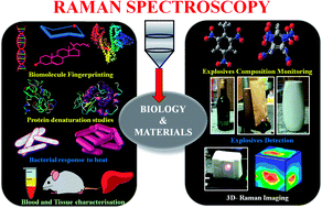
Raman spectroscopy has become an essential tool for chemists, physicists, biologists and materials scientists. In this article, we present the challenges in unravelling the molecule-specific Raman spectral signatures of different biomolecules like proteins, nucleic acids, lipids and carbohydrates based on the review of our work and the current trends in these areas. We also show how Raman spectroscopy can be used to probe the secondary and tertiary structural changes occurring during thermal denaturation of protein and lysozyme as well as more complex biological systems like bacteria. Complex biological systems like tissues, cells, blood serum etc. are also made up of such biomolecules. Using mice liver and blood serum, it is shown that different tissues yield their unique signature Raman spectra, owing to a difference in the relative composition of the biomolecules. Additionally, recent progress in Raman spectroscopy for diagnosing a multitude of diseases ranging from cancer to infection is also presented. The second part of this article focuses on applications of Raman spectroscopy to materials. As a first example, Raman spectroscopy of a melt cast explosives formulation was carried out to monitor the changes in the peaks which indicates the potential of this technique for remote process monitoring. The second example presents various modern methods of Raman spectroscopy such as spatially offset Raman spectroscopy (SORS), reflection, transmission and universal multiple angle Raman spectroscopy (UMARS) to study layered materials. Studies on chemicals/layered materials hidden in non-metallic containers using the above variants are presented. Using suitable examples, it is shown how a specific excitation or collection geometry can yield different information about the location of materials. Additionally, it is shown that UMARS imaging can also be used as an effective tool to obtain layer specific information of materials located at depths beyond a few centimeters.
中文翻译:

拉曼光谱在生物学和材料中的应用面临的挑战
拉曼光谱已成为化学家、物理学家、生物学家和材料科学家必不可少的工具。在本文中,我们基于对我们工作的回顾和这些领域的当前趋势,提出了解开不同生物分子(如蛋白质、核酸、脂质和碳水化合物)的分子特异性拉曼光谱特征的挑战。我们还展示了如何使用拉曼光谱来探测蛋白质和溶菌酶以及更复杂的生物系统(如细菌)热变性过程中发生的二级和三级结构变化。复杂的生物系统,如组织、细胞、血清等。也是由这样的生物分子组成的。使用小鼠肝脏和血清表明,由于生物分子的相对组成不同,不同的组织会产生其独特的拉曼光谱特征。此外,还介绍了拉曼光谱在诊断从癌症到感染等多种疾病方面的最新进展。本文的第二部分重点介绍拉曼光谱在材料中的应用。作为第一个例子,对熔铸炸药配方进行拉曼光谱以监测峰的变化,这表明该技术在远程过程监测中的潜力。第二个例子介绍了各种现代拉曼光谱方法,例如空间偏移拉曼光谱 (SORS)、反射、透射和通用多角度拉曼光谱 (UMARS) 来研究层状材料。介绍了使用上述变体对隐藏在非金属容器中的化学品/分层材料的研究。使用合适的示例,展示了特定的激发或收集几何形状如何产生有关材料位置的不同信息。此外,研究表明,UMARS 成像还可用作获取深度超过几厘米的材料的特定层信息的有效工具。
更新日期:2018-07-20
中文翻译:

拉曼光谱在生物学和材料中的应用面临的挑战
拉曼光谱已成为化学家、物理学家、生物学家和材料科学家必不可少的工具。在本文中,我们基于对我们工作的回顾和这些领域的当前趋势,提出了解开不同生物分子(如蛋白质、核酸、脂质和碳水化合物)的分子特异性拉曼光谱特征的挑战。我们还展示了如何使用拉曼光谱来探测蛋白质和溶菌酶以及更复杂的生物系统(如细菌)热变性过程中发生的二级和三级结构变化。复杂的生物系统,如组织、细胞、血清等。也是由这样的生物分子组成的。使用小鼠肝脏和血清表明,由于生物分子的相对组成不同,不同的组织会产生其独特的拉曼光谱特征。此外,还介绍了拉曼光谱在诊断从癌症到感染等多种疾病方面的最新进展。本文的第二部分重点介绍拉曼光谱在材料中的应用。作为第一个例子,对熔铸炸药配方进行拉曼光谱以监测峰的变化,这表明该技术在远程过程监测中的潜力。第二个例子介绍了各种现代拉曼光谱方法,例如空间偏移拉曼光谱 (SORS)、反射、透射和通用多角度拉曼光谱 (UMARS) 来研究层状材料。介绍了使用上述变体对隐藏在非金属容器中的化学品/分层材料的研究。使用合适的示例,展示了特定的激发或收集几何形状如何产生有关材料位置的不同信息。此外,研究表明,UMARS 成像还可用作获取深度超过几厘米的材料的特定层信息的有效工具。


 京公网安备 11010802027423号