当前位置:
X-MOL 学术
›
Nano-Micro Lett.
›
论文详情
Our official English website, www.x-mol.net, welcomes your
feedback! (Note: you will need to create a separate account there.)
In Vivo Tumor-Targeted Dual-Modality PET/Optical Imaging with a Yolk/Shell-Structured Silica Nanosystem
Nano-Micro Letters ( IF 31.6 ) Pub Date : 2018-07-16 , DOI: 10.1007/s40820-018-0216-2 Sixiang Shi , Feng Chen , Shreya Goel , Stephen A. Graves , Haiming Luo , Charles P. Theuer , Jonathan W. Engle , Weibo Cai
中文翻译:

卵黄/壳结构二氧化硅纳米系统体内靶向肿瘤的双峰PET /光学成像。
更新日期:2018-07-16
Nano-Micro Letters ( IF 31.6 ) Pub Date : 2018-07-16 , DOI: 10.1007/s40820-018-0216-2 Sixiang Shi , Feng Chen , Shreya Goel , Stephen A. Graves , Haiming Luo , Charles P. Theuer , Jonathan W. Engle , Weibo Cai

|
Silica nanoparticles have been one of the most promising nanosystems for biomedical applications due to their facile surface chemistry and non-toxic nature. However, it is still challenging to effectively deliver them into tumor sites and noninvasively visualize their in vivo biodistribution with excellent sensitivity and accuracy for effective cancer diagnosis. In this study, we design a yolk/shell-structured silica nanosystem 64Cu-NOTA-QD@HMSN-PEG-TRC105, which can be employed for tumor vasculature targeting and dual-modality PET/optical imaging, leading to superior targeting specificity, excellent imaging capability and more reliable diagnostic outcomes. By combining vasculature targeting, pH-sensitive drug delivery, and dual-modality imaging into a single platform, as-designed yolk/shell-structured silica nanosystems may be employed for the future image-guided tumor-targeted drug delivery, to further enable cancer theranostics.
Open image in new window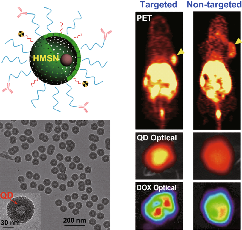
中文翻译:
卵黄/壳结构二氧化硅纳米系统体内靶向肿瘤的双峰PET /光学成像。
二氧化硅纳米粒子由于其易用的表面化学性质和无毒性质,已成为生物医学应用中最有前途的纳米系统之一。然而,将其有效地递送到肿瘤部位并以优异的灵敏度和准确性无创地观察其体内生物分布对于有效的癌症诊断仍然是挑战。在这项研究中,我们设计了一个卵黄/壳结构的二氧化硅纳米系统64Cu-NOTA-QD @ HMSN-PEG-TRC105可用于肿瘤血管靶向和双模式PET /光学成像,从而具有出色的靶向特异性,出色的成像能力和更可靠的诊断结果。通过将血管靶向,pH敏感药物递送和双峰成像结合到一个平台中,可以将设计好的卵黄/壳结构二氧化硅纳米系统用于未来的图像引导肿瘤靶向药物递送,以进一步实现癌症治疗学。
在新窗口中打开图像


 京公网安备 11010802027423号