当前位置:
X-MOL 学术
›
ChemCatChem
›
论文详情
Our official English website, www.x-mol.net, welcomes your feedback! (Note: you will need to create a separate account there.)
Nanoscale Chemical Imaging of a Single Catalyst Particle with Tip‐Enhanced Fluorescence Microscopy
ChemCatChem ( IF 4.5 ) Pub Date : 2018-07-31 , DOI: 10.1002/cctc.201801023 Naresh Kumar 1, 2 , Sam Kalirai 1 , Andrew J Wain 2 , Bert M Weckhuysen 1
ChemCatChem ( IF 4.5 ) Pub Date : 2018-07-31 , DOI: 10.1002/cctc.201801023 Naresh Kumar 1, 2 , Sam Kalirai 1 , Andrew J Wain 2 , Bert M Weckhuysen 1
Affiliation
|
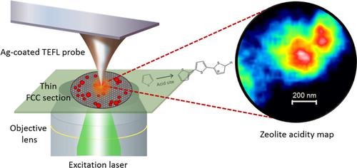
Determining the active site in real‐life solid catalysts remains an intellectual challenge and is crucial for exploring the road towards their rational design. In recent years various micro‐spectroscopic methods have revealed valuable structure‐activity data at the level of a single catalyst particle, even under reaction conditions. Herein, we introduce Tip‐Enhanced FLuorescence (TEFL) microscopy as a novel and versatile characterization tool for catalysis research. This has been achieved using a Fluid Catalytic Cracking (FCC) catalyst as showcase material. Thin sectioning of industrially used FCC particles together with selective staining of Brønsted acidity has enabled high‐resolution TEFL mapping of different catalyst regions. Hyperspectral information gained via TEFL microscopy reveals a spatial distribution of Brønsted acidity within individual zeolite domains in different regions of the FCC catalyst particle. Comparison of TEFL measurements from different FCC particles showed significant intra‐ and inter‐particle heterogeneities both in zeolite domain size and chemical reactivity.
中文翻译:

使用尖端增强荧光显微镜对单个催化剂颗粒进行纳米级化学成像
确定现实固体催化剂中的活性位点仍然是一项智力挑战,对于探索合理设计之路至关重要。近年来,各种显微光谱方法揭示了单个催化剂颗粒水平上的有价值的结构活性数据,甚至在反应条件下也是如此。在此,我们介绍尖端增强荧光(TEFL)显微镜作为催化研究的一种新颖且多功能的表征工具。这是通过使用流体催化裂化 (FCC) 催化剂作为展示材料来实现的。工业使用的 FCC 颗粒的薄切片以及布朗斯台德酸度的选择性染色使得不同催化剂区域的高分辨率 TEFL 绘图成为可能。通过 TEFL 显微镜获得的高光谱信息揭示了 FCC 催化剂颗粒不同区域的各个沸石域内布朗斯台德酸度的空间分布。不同 FCC 颗粒的 TEFL 测量结果的比较表明,在沸石域尺寸和化学反应性方面均存在显着的颗粒内和颗粒间异质性。
更新日期:2018-07-31
中文翻译:
使用尖端增强荧光显微镜对单个催化剂颗粒进行纳米级化学成像
确定现实固体催化剂中的活性位点仍然是一项智力挑战,对于探索合理设计之路至关重要。近年来,各种显微光谱方法揭示了单个催化剂颗粒水平上的有价值的结构活性数据,甚至在反应条件下也是如此。在此,我们介绍尖端增强荧光(TEFL)显微镜作为催化研究的一种新颖且多功能的表征工具。这是通过使用流体催化裂化 (FCC) 催化剂作为展示材料来实现的。工业使用的 FCC 颗粒的薄切片以及布朗斯台德酸度的选择性染色使得不同催化剂区域的高分辨率 TEFL 绘图成为可能。通过 TEFL 显微镜获得的高光谱信息揭示了 FCC 催化剂颗粒不同区域的各个沸石域内布朗斯台德酸度的空间分布。不同 FCC 颗粒的 TEFL 测量结果的比较表明,在沸石域尺寸和化学反应性方面均存在显着的颗粒内和颗粒间异质性。


 京公网安备 11010802027423号