Our official English website, www.x-mol.net, welcomes your feedback! (Note: you will need to create a separate account there.)
Necessary Experimental Conditions for Single-Shot Diffraction Imaging of DNA-Based Structures with X-ray Free-Electron Lasers
ACS Nano ( IF 17.1 ) Pub Date : 2018-07-09 00:00:00 , DOI: 10.1021/acsnano.8b01838 Zhibin Sun 1, 2, 3 , Jiadong Fan 3 , Haoyuan Li 2, 4 , Huajie Liu 5 , Daewoong Nam 6, 7 , Chan Kim 8, 9 , Yoonhee Kim 8, 9 , Yubo Han 10, 11 , Jianhua Zhang 1, 3 , Shengkun Yao 3 , Jaehyun Park 6 , Sunam Kim 6 , Kensuke Tono 12 , Makina Yabashi 13 , Tetsuya Ishikawa 13 , Changyong Song 7 , Chunhai Fan 5 , Huaidong Jiang 3
ACS Nano ( IF 17.1 ) Pub Date : 2018-07-09 00:00:00 , DOI: 10.1021/acsnano.8b01838 Zhibin Sun 1, 2, 3 , Jiadong Fan 3 , Haoyuan Li 2, 4 , Huajie Liu 5 , Daewoong Nam 6, 7 , Chan Kim 8, 9 , Yoonhee Kim 8, 9 , Yubo Han 10, 11 , Jianhua Zhang 1, 3 , Shengkun Yao 3 , Jaehyun Park 6 , Sunam Kim 6 , Kensuke Tono 12 , Makina Yabashi 13 , Tetsuya Ishikawa 13 , Changyong Song 7 , Chunhai Fan 5 , Huaidong Jiang 3
Affiliation

|
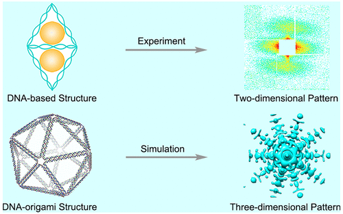
It has been proposed that the radiation damage to biological particles and soft condensed matter can be overcome by ultrafast and ultraintense X-ray free-electron lasers (FELs) with short pulse durations. The successful demonstration of the “diffraction-before-destruction” concept has made single-shot diffraction imaging a promising tool to achieve high resolutions under the native states of samples. However, the resolution is still limited because of the low signal-to-noise ratio, especially for biological specimens such as cells, viruses, and macromolecular particles. Here, we present a demonstration single-shot diffraction imaging experiment of DNA-based structures at SPring-8 Angstrom Compact Free Electron Laser (SACLA), Japan. Through quantitative analysis of the reconstructed images, the scattering abilities of gold and DNA were demonstrated. Suggestions for extracting valid DNA signals from noisy diffraction patterns were also explained and outlined. To sketch out the necessary experimental conditions for the 3D imaging of DNA origami or DNA macromolecular particles, we carried out numerical simulations with practical detector noise and experimental geometry using the Linac Coherent Light Source (LCLS) at the SLAC National Accelerator Laboratory, USA. The simulated results demonstrate that it is possible to capture images of DNA-based structures at high resolutions with the technique development of current and next-generation X-ray FEL facilities.
中文翻译:

X射线自由电子激光器对基于DNA的结构进行单次衍射成像的必要实验条件
已经提出,可以通过短脉冲持续时间的超快和超强X射线自由电子激光器(FEL)来克服对生物颗粒和软凝结物的辐射损害。“销毁前衍射”概念的成功演示使单次衍射成像成为在样品的原始状态下实现高分辨率的有前途的工具。但是,由于信噪比低,分辨率仍然受到限制,尤其是对于诸如细胞,病毒和大分子颗粒等生物标本而言。在这里,我们展示了日本SPring-8埃克斯紧凑型自由电子激光(SACLA)上基于DNA的结构的演示单次衍射成像实验。通过对重建图像的定量分析,证明了金和DNA的散射能力。还解释并概述了从噪声衍射图中提取有效DNA信号的建议。为了勾勒出DNA折纸或DNA大分子颗粒3D成像的必要实验条件,我们在美国SLAC国家加速器实验室使用Linac相干光源(LCLS)进行了具有实际检测器噪声和实验几何形状的数值模拟。仿真结果表明,随着当前和下一代X射线FEL设施的技术发展,有可能以高分辨率捕获基于DNA的结构的图像。我们在美国SLAC国家加速器实验室使用直线加速器相干光源(LCLS)进行了具有实际探测器噪声和实验几何形状的数值模拟。仿真结果表明,随着当前和下一代X射线FEL设施的技术发展,有可能以高分辨率捕获基于DNA的结构的图像。我们在美国SLAC国家加速器实验室使用直线加速器相干光源(LCLS)进行了具有实际探测器噪声和实验几何形状的数值模拟。仿真结果表明,随着当前和下一代X射线FEL设施的技术发展,有可能以高分辨率捕获基于DNA的结构的图像。
更新日期:2018-07-09
中文翻译:

X射线自由电子激光器对基于DNA的结构进行单次衍射成像的必要实验条件
已经提出,可以通过短脉冲持续时间的超快和超强X射线自由电子激光器(FEL)来克服对生物颗粒和软凝结物的辐射损害。“销毁前衍射”概念的成功演示使单次衍射成像成为在样品的原始状态下实现高分辨率的有前途的工具。但是,由于信噪比低,分辨率仍然受到限制,尤其是对于诸如细胞,病毒和大分子颗粒等生物标本而言。在这里,我们展示了日本SPring-8埃克斯紧凑型自由电子激光(SACLA)上基于DNA的结构的演示单次衍射成像实验。通过对重建图像的定量分析,证明了金和DNA的散射能力。还解释并概述了从噪声衍射图中提取有效DNA信号的建议。为了勾勒出DNA折纸或DNA大分子颗粒3D成像的必要实验条件,我们在美国SLAC国家加速器实验室使用Linac相干光源(LCLS)进行了具有实际检测器噪声和实验几何形状的数值模拟。仿真结果表明,随着当前和下一代X射线FEL设施的技术发展,有可能以高分辨率捕获基于DNA的结构的图像。我们在美国SLAC国家加速器实验室使用直线加速器相干光源(LCLS)进行了具有实际探测器噪声和实验几何形状的数值模拟。仿真结果表明,随着当前和下一代X射线FEL设施的技术发展,有可能以高分辨率捕获基于DNA的结构的图像。我们在美国SLAC国家加速器实验室使用直线加速器相干光源(LCLS)进行了具有实际探测器噪声和实验几何形状的数值模拟。仿真结果表明,随着当前和下一代X射线FEL设施的技术发展,有可能以高分辨率捕获基于DNA的结构的图像。


 京公网安备 11010802027423号