当前位置:
X-MOL 学术
›
Metallomics
›
论文详情
Our official English website, www.x-mol.net, welcomes your
feedback! (Note: you will need to create a separate account there.)
Selenoprotein-U (SelU) knockdown triggers autophagy through PI3K–Akt–mTOR pathway inhibition in rooster Sertoli cells
Metallomics ( IF 2.9 ) Pub Date : 2018-06-26 00:00:00 , DOI: 10.1039/c8mt00090e Hamid Sattar 1, 2, 3, 4 , Jie Yang 1, 2, 3, 4 , Xia Zhao 1, 2, 3, 4 , Jingzeng Cai 1, 2, 3, 4 , Qi Liu 1, 2, 3, 4 , Muhammad Ishfaq 1, 2, 3, 4 , Zijiang Yang 1, 2, 3, 4 , Menghao Chen 1, 2, 3, 4 , Ziwei Zhang 1, 2, 3, 4 , Shiwen Xu 1, 2, 3, 4
Metallomics ( IF 2.9 ) Pub Date : 2018-06-26 00:00:00 , DOI: 10.1039/c8mt00090e Hamid Sattar 1, 2, 3, 4 , Jie Yang 1, 2, 3, 4 , Xia Zhao 1, 2, 3, 4 , Jingzeng Cai 1, 2, 3, 4 , Qi Liu 1, 2, 3, 4 , Muhammad Ishfaq 1, 2, 3, 4 , Zijiang Yang 1, 2, 3, 4 , Menghao Chen 1, 2, 3, 4 , Ziwei Zhang 1, 2, 3, 4 , Shiwen Xu 1, 2, 3, 4
Affiliation
|
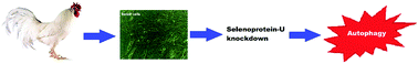
Selenium (Se) is a major component of male reproduction which exerts its effects via selenoproteins. Selenoprotein U (SelU), a newly identified protein, is expressed highly in eukaryotes and possesses a conserved motif similar to that existing in other thiol-dependent redox regulating selenoproteins; however its function is unknown. To investigate the role of SelU in testis autophagic and/or apoptosis cell death mechanisms, we established a Sertoli cell (SC) model isolated from 45 day old layer roosters. Small interfering RNA (siRNA) technology was used to develop SelU-knockdown (SelU-KD) and normal (N) SC models. Consequent to transfection, electron microscopy, qPCR, and western blot were performed. The results show that the mRNA and proteins of autophagy and anti-apoptosis genes increased while that of anti-autophagic mammalian target of rapamycin (mTOR) and pro-apoptosis genes decreased significantly in SelU-KD in contrast to N cells. Simultaneously, in contrast to N cells the expression of phosphoinositide-3-kinase (PI3K) and protein kinase B (PKB/Akt) both at the mRNA and protein levels decreased significantly in SelU-KD cells. In-addition, SelU depletion altered the expression of regulatory factors and increased the mRNA of TSC (tuberous sclerosis complex) genes as compared to N cells. Extensive autophagosome formation and lysosome degradation with an intact cytoskeleton were observed in SelU-KD cells. Our data indicate that SelU deprivation elicits autophagy and reduces the expression of important growth factors in SCs by disrupting the PI3K–Akt–mTOR signaling pathway. However SelU attenuation did not induce apoptosis in rooster SCs. Taken together, we conclude that SelU is essential for the survival and normal functioning of SCs.
中文翻译:

Selenoprotein-U(SelU)敲低可通过抑制PI3K–Akt–mTOR途径抑制雄性Sertoli细胞中的自噬
硒是男性生殖的主要成分,它通过以下方式发挥作用硒蛋白。硒蛋白U(SelU)是一种新近鉴定的蛋白,在真核生物中高度表达,并具有与其他巯基依赖性氧化还原调节硒蛋白相似的保守基序。但是其功能未知。为了研究SelU在睾丸自噬和/或凋亡细胞死亡机制中的作用,我们建立了一个从45天大的公鸡身上分离的Sertoli细胞(SC)模型。小干扰RNA(siRNA)技术用于开发SelU组合(SelU-KD)和正常(N)SC模型。转染的结果是,进行了电子显微镜,qPCR和蛋白质印迹。结果表明,与N细胞相比,SelU-KD中自噬和抗凋亡基因的mRNA和蛋白质增加,而雷帕霉素(mTOR)的抗自噬哺乳动物靶标和促凋亡基因的mRNA和蛋白质显着减少。同时,与N细胞相反,SelU-KD细胞中mRNA和蛋白水平的磷酸肌醇3激酶(PI3K)和蛋白激酶B(PKB / Akt)的表达均显着下降。此外,与N细胞相比,SelU耗竭改变了调节因子的表达并增加了TSC(结节性硬化复合物)基因的mRNA。在SelU-KD细胞中观察到广泛的自噬体形成和具有完整细胞骨架的溶酶体降解。我们的数据表明,SelU剥夺通过破坏PI3K–Akt–mTOR信号传导途径引起自噬,并降低SC中重要生长因子的表达。但是,SelU衰减不会诱导公鸡SC中的细胞凋亡。综上所述,我们得出结论,SelU对于SC的生存和正常运转至关重要。
更新日期:2018-06-26
中文翻译:
Selenoprotein-U(SelU)敲低可通过抑制PI3K–Akt–mTOR途径抑制雄性Sertoli细胞中的自噬
硒是男性生殖的主要成分,它通过以下方式发挥作用硒蛋白。硒蛋白U(SelU)是一种新近鉴定的蛋白,在真核生物中高度表达,并具有与其他巯基依赖性氧化还原调节硒蛋白相似的保守基序。但是其功能未知。为了研究SelU在睾丸自噬和/或凋亡细胞死亡机制中的作用,我们建立了一个从45天大的公鸡身上分离的Sertoli细胞(SC)模型。小干扰RNA(siRNA)技术用于开发SelU组合(SelU-KD)和正常(N)SC模型。转染的结果是,进行了电子显微镜,qPCR和蛋白质印迹。结果表明,与N细胞相比,SelU-KD中自噬和抗凋亡基因的mRNA和蛋白质增加,而雷帕霉素(mTOR)的抗自噬哺乳动物靶标和促凋亡基因的mRNA和蛋白质显着减少。同时,与N细胞相反,SelU-KD细胞中mRNA和蛋白水平的磷酸肌醇3激酶(PI3K)和蛋白激酶B(PKB / Akt)的表达均显着下降。此外,与N细胞相比,SelU耗竭改变了调节因子的表达并增加了TSC(结节性硬化复合物)基因的mRNA。在SelU-KD细胞中观察到广泛的自噬体形成和具有完整细胞骨架的溶酶体降解。我们的数据表明,SelU剥夺通过破坏PI3K–Akt–mTOR信号传导途径引起自噬,并降低SC中重要生长因子的表达。但是,SelU衰减不会诱导公鸡SC中的细胞凋亡。综上所述,我们得出结论,SelU对于SC的生存和正常运转至关重要。


 京公网安备 11010802027423号