当前位置:
X-MOL 学术
›
ACS Biomater. Sci. Eng.
›
论文详情
Our official English website, www.x-mol.net, welcomes your
feedback! (Note: you will need to create a separate account there.)
Collagen Fiber Orientation Regulates 3D Vascular Network Formation and Alignment
ACS Biomaterials Science & Engineering ( IF 5.4 ) Pub Date : 2018-06-26 00:00:00 , DOI: 10.1021/acsbiomaterials.8b00384 Michael G. McCoy 1 , Jane M. Wei 1, 2 , Siyoung Choi 1 , Julian Palacios Goerger 1 , Warren Zipfel 1 , Claudia Fischbach 1, 3
ACS Biomaterials Science & Engineering ( IF 5.4 ) Pub Date : 2018-06-26 00:00:00 , DOI: 10.1021/acsbiomaterials.8b00384 Michael G. McCoy 1 , Jane M. Wei 1, 2 , Siyoung Choi 1 , Julian Palacios Goerger 1 , Warren Zipfel 1 , Claudia Fischbach 1, 3
Affiliation

|
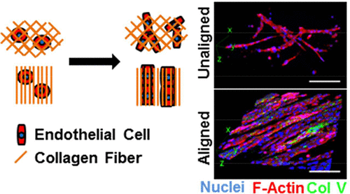
Alignment of collagen type I fibers is a hallmark of both physiological and pathological tissue remodeling. However, the effects of collagen fiber orientation on endothelial cell behavior and vascular network formation are poorly understood because of a lack of model systems that allow studying these potential functional connections. By casting collagen type I into prestrained (0, 10, 25, 50% strain), poly(dimethylsiloxane) (PDMS)-based microwells and releasing the mold strain following polymerization, we have created collagen gels with varying fiber alignment as confirmed by structural analysis. Endothelial cells embedded within the different gels responded to increased collagen fiber orientation by assembling into 3D vascular networks that consisted of thicker, more aligned branches and featured elevated collagen IV deposition and lumen formation relative to control conditions. These substrate-dependent changes in microvascular network formation were associated with altered cell division and migration patterns and related to enhanced mechanosignaling. Our studies indicate that collagen fiber alignment can directly regulate vascular network formation and that culture models with aligned collagen may be used to investigate the underlying mechanisms ultimately advancing our understanding of tissue development, homeostasis, and disease.
中文翻译:

胶原纤维的取向调节3D血管网络的形成和排列
I型胶原纤维的排列是生理和病理组织重塑的标志。然而,由于缺乏能够研究这些潜在功能连接的模型系统,人们对胶原纤维取向对内皮细胞行为和血管网络形成的影响了解甚少。通过将I型胶原蛋白注入预应变(0%,10%,25%,50%应变),基于聚二甲基硅氧烷(PDMS)的微孔中,并在聚合后释放模具应变,我们通过结构确定了具有不同纤维排列的胶原蛋白凝胶分析。嵌入不同凝胶中的内皮细胞通过组装成3D血管网络来响应增加的胶原纤维定向,该3D血管网络由较厚的,相对于对照条件,分支排列更紧密,胶原IV沉积和管腔形成增加。这些依赖于底物的微血管网络形成的变化与细胞分裂和迁移模式的改变有关,并与增强的机械信号传递有关。我们的研究表明,胶原纤维的排列可以直接调节血管网络的形成,具有排列好的胶原的培养模型可用于研究潜在的机制,最终增进我们对组织发育,体内平衡和疾病的理解。
更新日期:2018-06-26
中文翻译:

胶原纤维的取向调节3D血管网络的形成和排列
I型胶原纤维的排列是生理和病理组织重塑的标志。然而,由于缺乏能够研究这些潜在功能连接的模型系统,人们对胶原纤维取向对内皮细胞行为和血管网络形成的影响了解甚少。通过将I型胶原蛋白注入预应变(0%,10%,25%,50%应变),基于聚二甲基硅氧烷(PDMS)的微孔中,并在聚合后释放模具应变,我们通过结构确定了具有不同纤维排列的胶原蛋白凝胶分析。嵌入不同凝胶中的内皮细胞通过组装成3D血管网络来响应增加的胶原纤维定向,该3D血管网络由较厚的,相对于对照条件,分支排列更紧密,胶原IV沉积和管腔形成增加。这些依赖于底物的微血管网络形成的变化与细胞分裂和迁移模式的改变有关,并与增强的机械信号传递有关。我们的研究表明,胶原纤维的排列可以直接调节血管网络的形成,具有排列好的胶原的培养模型可用于研究潜在的机制,最终增进我们对组织发育,体内平衡和疾病的理解。


 京公网安备 11010802027423号