当前位置:
X-MOL 学术
›
Metallomics
›
论文详情
Our official English website, www.x-mol.net, welcomes your
feedback! (Note: you will need to create a separate account there.)
Conditioned medium from overly excitatory primary astrocytes induced by La3+ increases apoptosis in primary neurons via upregulating the expression of NMDA receptors
Metallomics ( IF 2.9 ) Pub Date : 2018-06-21 00:00:00 , DOI: 10.1039/c8mt00056e Yaling Sun 1, 2, 3, 4, 5 , Jinghua Yang 1, 2, 3, 4, 5 , Xiaoyu Hu 1, 2, 3, 4, 5 , Xiang Gao 1, 2, 3, 4, 5 , Yingqi Li 1, 2, 3, 4, 5 , Miao Yu 1, 2, 3, 4, 5 , Shiyu Liu 1, 2, 3, 4, 5 , Yanxin Lu 1, 2, 3, 4, 5 , Jing Wang 1, 2, 3, 4, 5 , Liling Huang 1, 2, 3, 4, 5 , Xiaobo Lu 1, 2, 3, 4, 5 , Cuihong Jin 1, 2, 3, 4, 5 , Shengwen Wu 1, 2, 3, 4, 5 , Yuan Cai 1, 2, 3, 4, 5
Metallomics ( IF 2.9 ) Pub Date : 2018-06-21 00:00:00 , DOI: 10.1039/c8mt00056e Yaling Sun 1, 2, 3, 4, 5 , Jinghua Yang 1, 2, 3, 4, 5 , Xiaoyu Hu 1, 2, 3, 4, 5 , Xiang Gao 1, 2, 3, 4, 5 , Yingqi Li 1, 2, 3, 4, 5 , Miao Yu 1, 2, 3, 4, 5 , Shiyu Liu 1, 2, 3, 4, 5 , Yanxin Lu 1, 2, 3, 4, 5 , Jing Wang 1, 2, 3, 4, 5 , Liling Huang 1, 2, 3, 4, 5 , Xiaobo Lu 1, 2, 3, 4, 5 , Cuihong Jin 1, 2, 3, 4, 5 , Shengwen Wu 1, 2, 3, 4, 5 , Yuan Cai 1, 2, 3, 4, 5
Affiliation
|
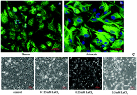
Lanthanum (La) can accumulate in the brain and impair learning and memory. However, the underlying mechanism of La-induced neurotoxicity has remained elusive. Under physiological conditions, it has been reported that moderately excitatory astrocytes play an important role in the regulation of neuronal signals and synaptic plasticity. However, under pathological conditions, overly excitatory astrocytes can release excess excitatory transmitters, such as glutamate (Glu) and D-serine, and induce the over-activation of NMDA receptors (NMDAR) in neurons, ultimately leading to neuronal excitotoxicity. To date, limited work has been performed with respect to whether La can induce neuronal excitotoxicity by inducing astrocytes to become overexcited. In this study, in vitro models of primary culture rat cortical astrocytes and neurons were established. First, the astrocytes were treated with 0.125 mM, 0.25 mM and 0.5 mM lanthanum chloride (LaCl3) for 24 h, and the supernatants were collected as a conditioned medium (CM) which is denoted as CM (La3+); then, the neurons were treated with CM (La3+) for 48 h. The results illustrate that LaCl3 treatment significantly upregulated the mRNA and protein expression levels of metabotropic glutamate receptor 5 (mGLUR5), phospholipase C (PLC), connexin 43 (Cx43) and Cx30, increased the concentrations of inositol trisphosphate (IP3) and [Ca2+]i, and promoted the synthesis and release of Glu and D-serine in astrocytes. Moreover, the CM (La3+) could increase the mRNA and protein expression levels of NMDAR subunits (NR1, NR2A, NR2B), the concentration of [Ca2+]i and the rate of apoptosis in neurons. Furthermore, after removal of La, CM (La-free) had a similar effect on neurons which could be antagonized by MK-801, DCKA and DAAO. These results suggest that the neuron apoptosis induced by La is closely related to the excessive release of Glu and D-serine from overly excitatory astrocytes.
中文翻译:

La 3+诱导的过度兴奋性原代星形胶质细胞的条件培养基通过上调NMDA受体的表达来增加原代神经元的凋亡
镧(La)可能在大脑中积聚并损害学习和记忆。但是,La引起的神经毒性的潜在机制仍然难以捉摸。据报道,在生理条件下,适度兴奋性星形胶质细胞在调节神经元信号和突触可塑性中起重要作用。但是,在病理条件下,过度兴奋的星形胶质细胞会释放过量的兴奋性递质,例如谷氨酸(Glu)和D-丝氨酸,并诱导神经元中NMDA受体(NMDAR)的过度活化,最终导致神经元兴奋性毒性。迄今为止,关于La是否可以通过诱导星形胶质细胞过度兴奋来诱导神经元兴奋性毒性,已经进行了有限的研究。在这项研究中,体外建立了原代培养大鼠皮质星形胶质细胞和神经元的模型。首先,将星形胶质细胞分别用0.125 mM,0.25 mM和0.5 mM氯化镧(LaCl 3)处理24 h,并收集上清液作为条件培养基(CM),表示为CM(La 3+);然后,用CM(La 3+)处理神经元48 h。结果表明,LaCl 3处理显着上调了促代谢型谷氨酸受体5(mGLUR 5),磷脂酶C(PLC),连接蛋白43(Cx43)和Cx30的mRNA和蛋白表达水平,增加了三磷酸肌醇(IP 3)和[Ca 2+ ] i,并促进星形胶质细胞中Glu和D-丝氨酸的合成和释放。此外,CM(La 3+)可以增加NMDAR亚基(NR1,NR2A,NR2B)的mRNA和蛋白表达水平,[Ca 2+ ] i的浓度以及神经元的凋亡率。此外,去除La后,CM(不含La)对神经元具有类似的作用,而MK-801,DCKA和DAAO可以拮抗。这些结果表明,La诱导的神经元凋亡与过度兴奋的星形胶质细胞中Glu和D-丝氨酸的过度释放密切相关。
更新日期:2018-06-21
中文翻译:
La 3+诱导的过度兴奋性原代星形胶质细胞的条件培养基通过上调NMDA受体的表达来增加原代神经元的凋亡
镧(La)可能在大脑中积聚并损害学习和记忆。但是,La引起的神经毒性的潜在机制仍然难以捉摸。据报道,在生理条件下,适度兴奋性星形胶质细胞在调节神经元信号和突触可塑性中起重要作用。但是,在病理条件下,过度兴奋的星形胶质细胞会释放过量的兴奋性递质,例如谷氨酸(Glu)和D-丝氨酸,并诱导神经元中NMDA受体(NMDAR)的过度活化,最终导致神经元兴奋性毒性。迄今为止,关于La是否可以通过诱导星形胶质细胞过度兴奋来诱导神经元兴奋性毒性,已经进行了有限的研究。在这项研究中,体外建立了原代培养大鼠皮质星形胶质细胞和神经元的模型。首先,将星形胶质细胞分别用0.125 mM,0.25 mM和0.5 mM氯化镧(LaCl 3)处理24 h,并收集上清液作为条件培养基(CM),表示为CM(La 3+);然后,用CM(La 3+)处理神经元48 h。结果表明,LaCl 3处理显着上调了促代谢型谷氨酸受体5(mGLUR 5),磷脂酶C(PLC),连接蛋白43(Cx43)和Cx30的mRNA和蛋白表达水平,增加了三磷酸肌醇(IP 3)和[Ca 2+ ] i,并促进星形胶质细胞中Glu和D-丝氨酸的合成和释放。此外,CM(La 3+)可以增加NMDAR亚基(NR1,NR2A,NR2B)的mRNA和蛋白表达水平,[Ca 2+ ] i的浓度以及神经元的凋亡率。此外,去除La后,CM(不含La)对神经元具有类似的作用,而MK-801,DCKA和DAAO可以拮抗。这些结果表明,La诱导的神经元凋亡与过度兴奋的星形胶质细胞中Glu和D-丝氨酸的过度释放密切相关。


 京公网安备 11010802027423号