Journal of the Mechanical Behavior of Biomedical Materials ( IF 3.3 ) Pub Date : 2018-06-18 , DOI: 10.1016/j.jmbbm.2018.06.022 S Oliviero 1 , M Giorgi 1 , E Dall'Ara 1
|
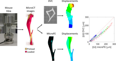
The mouse tibia is a common site to investigate bone adaptation. Micro-Finite Element (microFE) models based on micro-Computed Tomography (microCT) images can estimate bone mechanical properties non-invasively but their outputs need to be validated with experiments. Digital Volume Correlation (DVC) can provide experimental measurements of displacements over the whole bone volume. In this study we applied DVC to validate the local predictions of microFE models of the mouse tibia in compression.
Six mouse tibiae were stepwise compressed within a microCT system. MicroCT images were acquired in four configurations with applied compression of 0.5 N (preload), 6.5 N, 13.0 N and 19.5 N. Failure load was measured after the last scan. A global DVC algorithm was applied to the microCT images in order to obtain the displacement field over the bone volume. Homogeneous, isotropic linear hexahedral microFE models were generated from the images collected in the preload configuration with boundary conditions interpolated from the DVC displacements at the extremities of the tibia. Experimental displacements from DVC and numerical predictions were compared at corresponding locations in the middle of the bone. Stiffness and strength were also estimated from each model and compared with the experimental measurements.
The magnitude of the displacement vectors predicted by microFE models was highly correlated with experimental measurements (R2 >0.82). Higher but still reasonable errors were found for the Cartesian components. The models tended to overestimate local displacements in the longitudinal direction (R2 = 0.69–0.90, slope of the regression line=0.50–0.97). Errors in the prediction of structural mechanical properties were 14% ± 11% for stiffness and 9% ± 9% for strength.
In conclusion, the DVC approach has been applied to the validation of microFE models of the mouse tibia. The predictions of the models for both structural and local properties have been found reasonable for most preclinical applications.
中文翻译:
使用数字体积相关验证小鼠胫骨的有限元模型
小鼠胫骨是研究骨骼适应的常见部位。基于微计算机断层扫描 (microCT) 图像的微有限元 (microFE) 模型可以非侵入性地估计骨骼力学性能,但其输出需要通过实验进行验证。数字体积相关 (DVC) 可以提供对整个骨体积位移的实验测量。在这项研究中,我们应用 DVC 来验证小鼠胫骨受压的 microFE 模型的局部预测。
六只小鼠胫骨在 microCT 系统内逐步压缩。MicroCT 图像以四种配置获得,应用压缩为 0.5 N(预加载)、6.5 N、13.0 N 和 19.5 N。在最后一次扫描后测量失效载荷。为了获得骨体积上的位移场,将全局 DVC 算法应用于 microCT 图像。均匀的、各向同性的线性六面体 microFE 模型是从预加载配置中收集的图像生成的,边界条件从胫骨末端的 DVC 位移中插值。在骨骼中间的相应位置比较来自 DVC 的实验位移和数值预测。还从每个模型估计刚度和强度,并与实验测量值进行比较。
microFE 模型预测的位移矢量的大小与实验测量值高度相关(R 2 >0.82)。对于笛卡尔分量,发现了更高但仍然合理的错误。模型倾向于高估纵向的局部位移(R 2 = 0.69-0.90,回归线的斜率=0.50-0.97)。结构力学性能的预测误差为刚度的 14% ± 11% 和强度的 9% ± 9%。
总之,DVC 方法已应用于小鼠胫骨 microFE 模型的验证。对于大多数临床前应用,已发现模型对结构和局部特性的预测是合理的。


 京公网安备 11010802027423号