当前位置:
X-MOL 学术
›
Biochemistry
›
论文详情
Our official English website, www.x-mol.net, welcomes your
feedback! (Note: you will need to create a separate account there.)
X-ray Emission Spectroscopy as an in Situ Diagnostic Tool for X-ray Crystallography of Metalloproteins Using an X-ray Free-Electron Laser
Biochemistry ( IF 2.9 ) Pub Date : 2018-06-15 00:00:00 , DOI: 10.1021/acs.biochem.8b00325 Thomas Fransson 1 , Ruchira Chatterjee 2 , Franklin D. Fuller 3 , Sheraz Gul 2 , Clemens Weninger 3 , Dimosthenis Sokaras 4 , Thomas Kroll 4 , Roberto Alonso-Mori 3 , Uwe Bergmann 1 , Jan Kern 2 , Vittal K. Yachandra 2 , Junko Yano 2
Biochemistry ( IF 2.9 ) Pub Date : 2018-06-15 00:00:00 , DOI: 10.1021/acs.biochem.8b00325 Thomas Fransson 1 , Ruchira Chatterjee 2 , Franklin D. Fuller 3 , Sheraz Gul 2 , Clemens Weninger 3 , Dimosthenis Sokaras 4 , Thomas Kroll 4 , Roberto Alonso-Mori 3 , Uwe Bergmann 1 , Jan Kern 2 , Vittal K. Yachandra 2 , Junko Yano 2
Affiliation

|
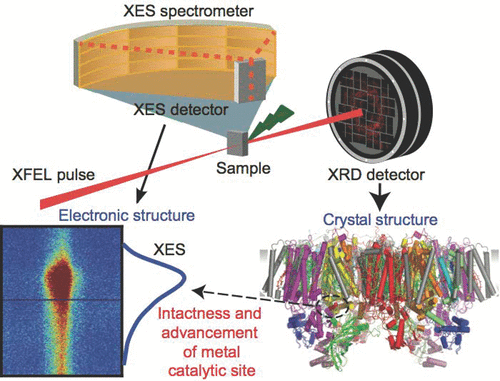
Serial femtosecond crystallography (SFX) using the ultrashort X-ray pulses from a X-ray free-electron laser (XFEL) provides a new way of collecting structural data at room temperature that allows for following the reaction in real time after initiation. XFEL experiments are conducted in a shot-by-shot mode as the sample is destroyed and replenished after each X-ray pulse, and therefore, monitoring and controlling the data quality by using in situ diagnostic tools is critical. To study metalloenzymes, we developed the use of simultaneous collection of X-ray diffraction of crystals along with X-ray emission spectroscopy (XES) data that is used as a diagnostic tool for crystallography, by monitoring the chemical state of the metal catalytic center. We have optimized data analysis methods and sample delivery techniques for fast and active feedback to ensure the quality of each batch of samples and the turnover of the catalytic reaction caused by reaction triggering methods. Here, we describe this active in situ feedback system using Photosystem II as an example that catalyzes the oxidation of H2O to O2 at the Mn4CaO5 active site. We used the first moments of the Mn Kβ1,3 emission spectra, which are sensitive to the oxidation state of Mn, as the primary diagnostics. This approach is applicable to different metalloproteins to determine the integrity of samples and follow changes in the chemical states of the reaction that can be initiated by light or activated by substrates and offers a metric for determining the diffraction images that are used for the final data sets.
中文翻译:

X射线发射光谱作为使用X射线自由电子激光对金属蛋白进行X射线晶体学分析的原位诊断工具
使用来自X射线自由电子激光器(XFEL)的超短X射线脉冲的连续飞秒晶体学(SFX)提供了一种在室温下收集结构数据的新方法,该方法可在引发后实时跟踪反应。XFEL实验是在每次X射线脉冲后销毁并补充样品的情况下,以逐次进行的方式进行的,因此,可以通过使用原位监测和控制数据质量诊断工具至关重要。为了研究金属酶,我们通过监测金属催化中心的化学状态,开发了同时收集晶体的X射线衍射和X射线发射光谱(XES)数据(用作结晶学诊断工具)的用途。我们已经优化了数据分析方法和样品传递技术,以实现快速,主动的反馈,以确保每批样品的质量以及由反应触发方法引起的催化反应的周转率。在这里,我们以光电系统II为例,描述这种主动的原位反馈系统,该系统在Mn 4 CaO 5上催化H 2 O氧化为O 2活动站点。我们使用的MnKβ的第一时刻1,3-发射光谱,其是与Mn的氧化态敏感,作为主要诊断。该方法适用于不同的金属蛋白,以确定样品的完整性并跟踪反应的化学状态变化,该变化可以由光引发或由底物激活,并提供确定最终数据集所用衍射图像的度量。
更新日期:2018-06-15
中文翻译:

X射线发射光谱作为使用X射线自由电子激光对金属蛋白进行X射线晶体学分析的原位诊断工具
使用来自X射线自由电子激光器(XFEL)的超短X射线脉冲的连续飞秒晶体学(SFX)提供了一种在室温下收集结构数据的新方法,该方法可在引发后实时跟踪反应。XFEL实验是在每次X射线脉冲后销毁并补充样品的情况下,以逐次进行的方式进行的,因此,可以通过使用原位监测和控制数据质量诊断工具至关重要。为了研究金属酶,我们通过监测金属催化中心的化学状态,开发了同时收集晶体的X射线衍射和X射线发射光谱(XES)数据(用作结晶学诊断工具)的用途。我们已经优化了数据分析方法和样品传递技术,以实现快速,主动的反馈,以确保每批样品的质量以及由反应触发方法引起的催化反应的周转率。在这里,我们以光电系统II为例,描述这种主动的原位反馈系统,该系统在Mn 4 CaO 5上催化H 2 O氧化为O 2活动站点。我们使用的MnKβ的第一时刻1,3-发射光谱,其是与Mn的氧化态敏感,作为主要诊断。该方法适用于不同的金属蛋白,以确定样品的完整性并跟踪反应的化学状态变化,该变化可以由光引发或由底物激活,并提供确定最终数据集所用衍射图像的度量。


 京公网安备 11010802027423号