当前位置:
X-MOL 学术
›
Mater. Chem. Front.
›
论文详情
Our official English website, www.x-mol.net, welcomes your
feedback! (Note: you will need to create a separate account there.)
AIE-based super-resolution imaging probes for β-amyloid plaques in mouse brains†
Materials Chemistry Frontiers ( IF 6.0 ) Pub Date : 2018-06-14 00:00:00 , DOI: 10.1039/c8qm00209f Ya-Long Wang 1, 2, 3, 4, 5 , Cheng Fan 1, 2, 3, 4, 5 , Bo Xin 1, 2, 3, 4, 5 , Jian-Ping Zhang 1, 2, 3, 4, 5 , Ting Luo 1, 2, 3, 4, 5 , Ze-Qiang Chen 1, 2, 3, 4, 5 , Qi-Yuan Zhou 1, 2, 3, 4, 5 , Qi Yu 1, 2, 3, 4, 5 , Xiang-Ning Li 1, 2, 3, 4, 5 , Zhen-Li Huang 1, 2, 3, 4, 5 , Chong Li 1, 2, 3, 4, 5 , Ming-Qiang Zhu 1, 2, 3, 4, 5 , Ben Zhong Tang 5, 6, 7, 8
Materials Chemistry Frontiers ( IF 6.0 ) Pub Date : 2018-06-14 00:00:00 , DOI: 10.1039/c8qm00209f Ya-Long Wang 1, 2, 3, 4, 5 , Cheng Fan 1, 2, 3, 4, 5 , Bo Xin 1, 2, 3, 4, 5 , Jian-Ping Zhang 1, 2, 3, 4, 5 , Ting Luo 1, 2, 3, 4, 5 , Ze-Qiang Chen 1, 2, 3, 4, 5 , Qi-Yuan Zhou 1, 2, 3, 4, 5 , Qi Yu 1, 2, 3, 4, 5 , Xiang-Ning Li 1, 2, 3, 4, 5 , Zhen-Li Huang 1, 2, 3, 4, 5 , Chong Li 1, 2, 3, 4, 5 , Ming-Qiang Zhu 1, 2, 3, 4, 5 , Ben Zhong Tang 5, 6, 7, 8
Affiliation
|
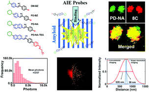
Fluorescence microscopy is an intuitive detection method of β-amyloid (Aβ) fibrillation, which usually occurs in early-stage Alzheimer's disease. With the aid of single-molecular localization of reversibly-activated fluorogens, the limit of optical diffraction in fluorescence microscopy can be overcome and the imaging resolution can be promoted to the sub-100 nm boundary. Aggregation-induced emission (AIE) is the fluorogenic emission behavior in which fluorescence is significantly enhanced due to the restriction of intramolecular motion when the fluorogens aggregate together or bind with specific targets. Reversible binding events of AIE-active fluorogens cause fluorescence switching enabling single-molecular localization and super-resolution imaging. Here, we report a series of super-resolution fluorescent probes with AIE activity, which are used for the detection and super-resolution imaging of fibrillar amyloids. The AIE-active fluorogens show superior in vitro sensitivity to fibrillar amyloids of hen egg white lysozyme (HEWL), which is usually used as a model protein for amyloid studies, with the limit of detection down to 63.71 nM. The fluorescence colocalization imaging indicates the excellent ex vivo targeting capability of Aβ plaques in mouse brain slices, with the colocalization degrees more than 90%. Based on the reversible binding between AIE-fluorogens and Aβ fibrils, the AIE-based super-resolution imaging of in vitro Aβ fibrillation in tubes and ex vivo Aβ plaques in mouse brain slices is accomplished. The detailed structure information reveals that Aβ plaques in the mouse brain are composed of numerous radiant nanofibrils with an optical imaging resolution of about 30 nm.
中文翻译:

基于AIE的超高分辨率成像探针,可用于小鼠大脑中的β-淀粉样斑块†
荧光显微镜检查是一种直观的检测β淀粉样蛋白(Aβ)颤动的方法,通常发生在早期阿尔茨海默氏病中。借助于可逆活化的氟原子的单分子定位,可以克服荧光显微镜中光学衍射的局限,并且可以将成像分辨率提高到100 nm以下。聚集诱导发射(AIE)是一种荧光发射行为,其中,当氟团聚在一起或与特定靶标结合时,由于分子内运动的限制,荧光显着增强。AIE活性氟的可逆结合事件导致荧光转换,从而实现单分子定位和超分辨率成像。在这里,我们报告了一系列具有AIE活性的超分辨率荧光探针,用于检测纤维状淀粉样蛋白和超分辨率成像。AIE活性氟显示出优异的性能卵清蛋白溶菌酶(HEWL)对原纤维淀粉样蛋白的体外敏感性,通常用作淀粉样蛋白研究的模型蛋白,检出限低至63.71 nM。荧光共定位成像表明小鼠脑切片中Aβ斑块具有出色的离体靶向能力,共定位度超过90%。基于AIE氟与Aβ原纤维之间的可逆结合,完成了基于AIE的管体外Aβ原纤化和小鼠脑切片中离体Aβ斑块的超分辨率成像。详细的结构信息显示,小鼠大脑中的Aβ斑块由许多辐射纳米纤维组成,其光学成像分辨率约为30 nm。
更新日期:2018-06-14
中文翻译:
基于AIE的超高分辨率成像探针,可用于小鼠大脑中的β-淀粉样斑块†
荧光显微镜检查是一种直观的检测β淀粉样蛋白(Aβ)颤动的方法,通常发生在早期阿尔茨海默氏病中。借助于可逆活化的氟原子的单分子定位,可以克服荧光显微镜中光学衍射的局限,并且可以将成像分辨率提高到100 nm以下。聚集诱导发射(AIE)是一种荧光发射行为,其中,当氟团聚在一起或与特定靶标结合时,由于分子内运动的限制,荧光显着增强。AIE活性氟的可逆结合事件导致荧光转换,从而实现单分子定位和超分辨率成像。在这里,我们报告了一系列具有AIE活性的超分辨率荧光探针,用于检测纤维状淀粉样蛋白和超分辨率成像。AIE活性氟显示出优异的性能卵清蛋白溶菌酶(HEWL)对原纤维淀粉样蛋白的体外敏感性,通常用作淀粉样蛋白研究的模型蛋白,检出限低至63.71 nM。荧光共定位成像表明小鼠脑切片中Aβ斑块具有出色的离体靶向能力,共定位度超过90%。基于AIE氟与Aβ原纤维之间的可逆结合,完成了基于AIE的管体外Aβ原纤化和小鼠脑切片中离体Aβ斑块的超分辨率成像。详细的结构信息显示,小鼠大脑中的Aβ斑块由许多辐射纳米纤维组成,其光学成像分辨率约为30 nm。


 京公网安备 11010802027423号