当前位置:
X-MOL 学术
›
J. Mater. Chem. B
›
论文详情
Our official English website, www.x-mol.net, welcomes your
feedback! (Note: you will need to create a separate account there.)
Potential detection of cancer with fluorinated silicon nanoparticles in 19F MR and fluorescence imaging†
Journal of Materials Chemistry B ( IF 6.1 ) Pub Date : 2018-05-31 00:00:00 , DOI: 10.1039/c8tb00648b Sha Li 1, 2, 3, 4, 5 , Yaping Yuan 1, 2, 3, 4, 5 , Yuqi Yang 1, 2, 3, 4, 5 , Conggang Li 1, 2, 3, 4, 5 , Michael T. McMahon 6, 7, 8, 9, 10 , Maili Liu 1, 2, 3, 4, 5 , Shizhen Chen 1, 2, 3, 4, 5 , Xin Zhou 1, 2, 3, 4, 5
Journal of Materials Chemistry B ( IF 6.1 ) Pub Date : 2018-05-31 00:00:00 , DOI: 10.1039/c8tb00648b Sha Li 1, 2, 3, 4, 5 , Yaping Yuan 1, 2, 3, 4, 5 , Yuqi Yang 1, 2, 3, 4, 5 , Conggang Li 1, 2, 3, 4, 5 , Michael T. McMahon 6, 7, 8, 9, 10 , Maili Liu 1, 2, 3, 4, 5 , Shizhen Chen 1, 2, 3, 4, 5 , Xin Zhou 1, 2, 3, 4, 5
Affiliation
|
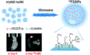
Fluorescence is widely used for cell imaging due to its high sensitivity and rich color choices but limited for in vivo imaging because of its low light penetration. Meanwhile, magnetic resonance imaging (MRI) is widely applied for in vivo diagnosis but not suitable for cell imaging because of its low resolution. As a result of rare background in living organisms, 19F-MRI stands out in several fields of clinical application. Herein, we report a one-pot microwave synthesis of fluorinated silicon nanoparticles (19FSiNPs), for detection of cancer cells and tumors. Based on the quantum effects of the nano-sized nanoparticles, 19FSiNPs can act as a label free dye for ultracontrast cell fluorescence imaging. Our experiments demonstrated that these nanoprobes significantly enhanced in vivo19F/1H MRI contrast in rats with non-small cell lung tumors. Moreover, the resulting 19FSiNPs exhibited high water dispersibility and excellent biocompatibility, which make them promising for both cell imaging and in vivo imaging applications.
中文翻译:

氟化硅纳米粒子在19 F MR和荧光成像中的潜在癌症检测能力†
荧光由于其高灵敏度和丰富的色彩选择而被广泛用于细胞成像,但由于其低的光穿透性而在体内成像中受到限制。同时,磁共振成像(MRI)由于其低分辨率而被广泛用于体内诊断,但不适用于细胞成像。由于活生物体中稀有的背景,19 F-MRI在临床应用的几个领域中脱颖而出。在这里,我们报告了氟化硅纳米颗粒(19 FSiNPs)的一锅微波合成,用于检测癌细胞和肿瘤。基于纳米粒子的量子效应,19FSiNP可充当无标记染料,用于超对比细胞荧光成像。我们的实验表明,这些纳米探针在患有非小细胞肺癌的大鼠中显着增强了体内19 F / 1 H MRI对比度。此外,所得的19种FSiNPs表现出高的水分散性和优异的生物相容性,这使其对于细胞成像和体内成像应用均充满希望。
更新日期:2018-05-31
中文翻译:
氟化硅纳米粒子在19 F MR和荧光成像中的潜在癌症检测能力†
荧光由于其高灵敏度和丰富的色彩选择而被广泛用于细胞成像,但由于其低的光穿透性而在体内成像中受到限制。同时,磁共振成像(MRI)由于其低分辨率而被广泛用于体内诊断,但不适用于细胞成像。由于活生物体中稀有的背景,19 F-MRI在临床应用的几个领域中脱颖而出。在这里,我们报告了氟化硅纳米颗粒(19 FSiNPs)的一锅微波合成,用于检测癌细胞和肿瘤。基于纳米粒子的量子效应,19FSiNP可充当无标记染料,用于超对比细胞荧光成像。我们的实验表明,这些纳米探针在患有非小细胞肺癌的大鼠中显着增强了体内19 F / 1 H MRI对比度。此外,所得的19种FSiNPs表现出高的水分散性和优异的生物相容性,这使其对于细胞成像和体内成像应用均充满希望。


 京公网安备 11010802027423号