当前位置:
X-MOL 学术
›
Anal. Methods
›
论文详情
Our official English website, www.x-mol.net, welcomes your
feedback! (Note: you will need to create a separate account there.)
Quantitation of acute monocytic leukemia cells spiked in control monocytes using surface-enhanced Raman spectroscopy
Analytical Methods ( IF 2.7 ) Pub Date : 2018-05-25 00:00:00 , DOI: 10.1039/c8ay01046c M. Hassoun 1, 2, 3, 4, 5 , N. Köse 3, 6, 7, 8 , R. Kiselev 1, 2, 3, 4, 5 , T. Kirchberger-Tolstik 1, 2, 3, 6, 7 , I. W. Schie 1, 2, 3 , C. Krafft 1, 2, 3 , J. Popp 1, 2, 3, 4, 5
Analytical Methods ( IF 2.7 ) Pub Date : 2018-05-25 00:00:00 , DOI: 10.1039/c8ay01046c M. Hassoun 1, 2, 3, 4, 5 , N. Köse 3, 6, 7, 8 , R. Kiselev 1, 2, 3, 4, 5 , T. Kirchberger-Tolstik 1, 2, 3, 6, 7 , I. W. Schie 1, 2, 3 , C. Krafft 1, 2, 3 , J. Popp 1, 2, 3, 4, 5
Affiliation

|
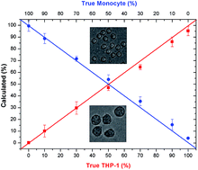
Surface enhanced Raman spectroscopy (SERS) was used to quantify leukemia cells spiked in control cells. The novelty of the technique lies in preparing cell lysates by ultrasound sonication and mixing with silver nanoparticles which allow reproducible interaction of biomolecules and nanoparticles. The SERS spectra of these mixtures not only exhibit enhanced bands of intracellular proteins and nucleic acids, but also spectral variations for accurate cell identification. Here, samples from an acute monocytic leukemia cell line, THP-1, and control monocytes from three donors served as the in vitro model system for leukemia. For quantitative analysis, seven mixtures containing different percentile amounts of leukemia lysates and lysates from control monocytes were prepared ranging from 0% to 100% and SERS spectra were measured. The more intense spectral contributions of proteins relative to nucleic acids correlated with the larger cytoplasm to nucleus ratio of leukemia cells than control monocytes. The experimental SERS spectra were fitted by a non-negative least squares (NNLS) algorithm to calculate the percentile amounts of each of the cell types and to determine their contributions to the mixtures. Even in a mixture with control monocytes (360 μl), a small amount (5 μl) of leukemia cells was detected, which represents 10% of leukemia cells considering their twofold larger diameter and eightfold larger volume. As this value is well below the threshold of 20% blast cells for leukemia diagnosis, this approach is very promising for both qualitative and quantitative analysis of human cell mixtures. This study demonstrates the potential of SERS and NNLS fitting as a rapid method for diagnosis of acute monocytic leukemia in human blood or bone marrow samples because only a single spectrum is required.
中文翻译:

使用表面增强拉曼光谱定量测定对照单核细胞中掺入的急性单核细胞白血病细胞
使用表面增强拉曼光谱(SERS)来量化加标在对照细胞中的白血病细胞。该技术的新颖之处在于通过超声处理并与银纳米颗粒混合制备细胞裂解物,从而使生物分子和纳米颗粒之间具有可重复的相互作用。这些混合物的SERS光谱不仅显示出增强的细胞内蛋白质和核酸谱带,而且还显示出光谱变化,以进行准确的细胞鉴定。在这里,来自急性单核细胞白血病细胞系THP-1和来自三个供体的对照单核细胞的样品用作体外白血病模型系统。为了进行定量分析,制备了七种含有不同百分数的白血病裂解物和对照单核细胞裂解物的混合物,并测量了SERS光谱。蛋白质相对于核酸的更强烈的光谱贡献与白血病细胞比对照单核细胞更大的细胞质与细胞核比有关。通过非负最小二乘(NNLS)算法拟合实验性SERS光谱,以计算每种细胞类型的百分位数,并确定它们对混合物的贡献。即使在与对照单核细胞(360μl)的混合物中,也检测到少量(5μl)的白血病细胞,考虑到它们的两倍大直径和八倍大体积,它们代表了白血病细胞的10%。由于该值远低于用于白血病诊断的20%原始细胞的阈值,因此该方法对于人细胞混合物的定性和定量分析都非常有前途。这项研究证明了SERS和NNLS拟合作为诊断人血或骨髓样品中急性单核细胞白血病的快速方法的潜力,因为仅需要一个光谱。
更新日期:2018-05-25
中文翻译:

使用表面增强拉曼光谱定量测定对照单核细胞中掺入的急性单核细胞白血病细胞
使用表面增强拉曼光谱(SERS)来量化加标在对照细胞中的白血病细胞。该技术的新颖之处在于通过超声处理并与银纳米颗粒混合制备细胞裂解物,从而使生物分子和纳米颗粒之间具有可重复的相互作用。这些混合物的SERS光谱不仅显示出增强的细胞内蛋白质和核酸谱带,而且还显示出光谱变化,以进行准确的细胞鉴定。在这里,来自急性单核细胞白血病细胞系THP-1和来自三个供体的对照单核细胞的样品用作体外白血病模型系统。为了进行定量分析,制备了七种含有不同百分数的白血病裂解物和对照单核细胞裂解物的混合物,并测量了SERS光谱。蛋白质相对于核酸的更强烈的光谱贡献与白血病细胞比对照单核细胞更大的细胞质与细胞核比有关。通过非负最小二乘(NNLS)算法拟合实验性SERS光谱,以计算每种细胞类型的百分位数,并确定它们对混合物的贡献。即使在与对照单核细胞(360μl)的混合物中,也检测到少量(5μl)的白血病细胞,考虑到它们的两倍大直径和八倍大体积,它们代表了白血病细胞的10%。由于该值远低于用于白血病诊断的20%原始细胞的阈值,因此该方法对于人细胞混合物的定性和定量分析都非常有前途。这项研究证明了SERS和NNLS拟合作为诊断人血或骨髓样品中急性单核细胞白血病的快速方法的潜力,因为仅需要一个光谱。


 京公网安备 11010802027423号