Journal of the Mechanical Behavior of Biomedical Materials ( IF 3.3 ) Pub Date : 2018-05-17 , DOI: 10.1016/j.jmbbm.2018.05.005 Marc A Stadelmann 1 , Ghislain Maquer 1 , Benjamin Voumard 1 , Aaron Grant 2 , David B Hackney 2 , Peter Vermathen 3 , Ron N Alkalay 4 , Philippe K Zysset 1
|
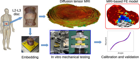
Intervertebral disc degeneration is a common disease that is often related to impaired mechanical function, herniations and chronic back pain. The degenerative process induces alterations of the disc's shape, composition and structure that can be visualized in vivo using magnetic resonance imaging (MRI). Numerical tools such as finite element analysis (FEA) have the potential to relate MRI-based information to the altered mechanical behavior of the disc. However, in terms of geometry, composition and fiber architecture, current FE models rely on observations made on healthy discs and might therefore not be well suited to study the degeneration process. To address the issue, we propose a new, more realistic FE methodology based on diffusion tensor imaging (DTI). For this study, a human disc joint was imaged in a high-field MR scanner with proton-density weighted (PD) and DTI sequences. The PD image was segmented and an anatomy-specific mesh was generated. Assuming accordance between local principal diffusion direction and local mean collagen fiber alignment, corresponding fiber angles were assigned to each element. Those element-wise fiber directions and PD intensities allowed the homogenized model to smoothly account for composition and fibrous structure of the disc. The disc's in vitro mechanical behavior was quantified under tension, compression, flexion, extension, lateral bending and rotation. The six resulting load-displacement curves could be replicated by the FE model, which supports our approach as a first proof of concept towards patient-specific disc modeling.
中文翻译:
在人椎间盘的有限元模型中集成基于 MRI 的几何结构、成分和纤维结构
椎间盘退变是一种常见病,常与机械功能受损、突出和慢性背痛有关。退行性过程会引起椎间盘形状、成分和结构的改变,这些改变可以在体内观察到使用磁共振成像 (MRI)。有限元分析 (FEA) 等数值工具有可能将基于 MRI 的信息与椎间盘改变的机械行为联系起来。然而,就几何、成分和纤维结构而言,当前的有限元模型依赖于对健康椎间盘的观察,因此可能不太适合研究退化过程。为了解决这个问题,我们提出了一种基于扩散张量成像(DTI)的新的、更现实的有限元方法。在这项研究中,人类椎间盘关节在具有质子密度加权 (PD) 和 DTI 序列的高场 MR 扫描仪中成像。对 PD 图像进行分割并生成特定解剖结构的网格。假设局部主扩散方向与局部平均胶原纤维排列一致,相应的纤维角度被分配给每个元素。这些元素方向的纤维方向和 PD 强度使均质模型能够平滑地解释椎间盘的成分和纤维结构。光盘的在张力、压缩、屈曲、伸展、横向弯曲和旋转下量化体外机械行为。FE 模型可以复制所产生的六个负载-位移曲线,这支持我们的方法作为针对患者特定椎间盘建模的第一个概念证明。


 京公网安备 11010802027423号