Analytical and Bioanalytical Chemistry ( IF 3.8 ) Pub Date : 2018-05-10 , DOI: 10.1007/s00216-018-1106-7 Seungkuk Ahn 1 , Herdeline Ann M Ardoña 1 , Johan U Lind 1, 2 , Feyisayo Eweje 1 , Sean L Kim 1 , Grant M Gonzalez 1 , Qihan Liu 1 , John F Zimmerman 1 , Georgios Pyrgiotakis 3 , Zhenyuan Zhang 3 , Juan Beltran-Huarac 3 , Paul Carpinone 3 , Brij M Moudgil 3 , Philip Demokritou 3 , Kevin Kit Parker 1
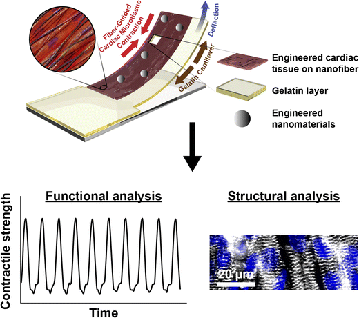
Due to the unique physicochemical properties exhibited by materials with nanoscale dimensions, there is currently a continuous increase in the number of engineered nanomaterials (ENMs) used in consumer goods. However, several reports associate ENM exposure to negative health outcomes such as cardiovascular diseases. Therefore, understanding the pathological consequences of ENM exposure represents an important challenge, requiring model systems that can provide mechanistic insights across different levels of ENM-based toxicity. To achieve this, we developed a mussel-inspired 3D microphysiological system (MPS) to measure cardiac contractility in the presence of ENMs. While multiple cardiac MPS have been reported as alternatives to in vivo testing, most systems only partially recapitulate the native extracellular matrix (ECM) structure. Here, we show how adhesive and aligned polydopamine (PDA)/polycaprolactone (PCL) nanofiber can be used to emulate the 3D native ECM environment of the myocardium. Such nanofiber scaffolds can support the formation of anisotropic and contractile muscular tissues. By integrating these fibers in a cardiac MPS, we assessed the effects of TiO2 and Ag nanoparticles on the contractile function of cardiac tissues. We found that these ENMs decrease the contractile function of cardiac tissues through structural damage to tissue architecture. Furthermore, the MPS with embedded sensors herein presents a way to non-invasively monitor the effects of ENM on cardiac tissue contractility at different time points. These results demonstrate the utility of our MPS as an analytical platform for understanding the functional impacts of ENMs while providing a biomimetic microenvironment to in vitro cardiac tissue samples.
Heart-on-a-chip integrated with mussel-inspired fiber scaffolds for a high-throughput toxicological assessment of engineered nanomaterials
中文翻译:
受贻贝启发的 3D 纤维支架,用于工程纳米材料的芯片心脏毒性研究
由于纳米级尺寸材料表现出独特的物理化学性质,目前消费品中使用的工程纳米材料(ENM)的数量不断增加。然而,一些报告将 ENM 暴露与心血管疾病等负面健康结果联系起来。因此,了解 ENM 暴露的病理后果是一个重要的挑战,需要模型系统能够提供不同级别的 ENM 毒性的机制见解。为了实现这一目标,我们开发了一种受贻贝启发的 3D 微生理系统 (MPS),用于测量 ENM 存在下的心肌收缩力。虽然多种心脏 MPS 已被报道为体内测试的替代方案,但大多数系统仅部分概括了天然细胞外基质 (ECM) 结构。在这里,我们展示了如何使用粘合且对齐的聚多巴胺 (PDA)/聚己内酯 (PCL) 纳米纤维来模拟心肌的 3D 原生 ECM 环境。这种纳米纤维支架可以支持各向异性和收缩性肌肉组织的形成。通过将这些纤维整合到心脏 MPS 中,我们评估了 TiO 2和 Ag 纳米颗粒对心脏组织收缩功能的影响。我们发现这些 ENM 通过对组织结构的结构损伤来降低心脏组织的收缩功能。此外,本文中具有嵌入式传感器的 MPS 提出了一种非侵入性监测 ENM 在不同时间点对心脏组织收缩性的影响的方法。这些结果证明了我们的 MPS 作为分析平台的实用性,可用于了解 ENM 的功能影响,同时为体外心脏组织样本提供仿生微环境。
在新窗口中打开图像
芯片上的心脏与受贻贝启发的纤维支架集成,用于工程纳米材料的高通量毒理学评估


 京公网安备 11010802027423号