当前位置:
X-MOL 学术
›
J. Anal. At. Spectrom.
›
论文详情
Our official English website, www.x-mol.net, welcomes your
feedback! (Note: you will need to create a separate account there.)
New applications of spectroscopy techniques reveal phylogenetically significant soft tissue residue in Paleozoic conodonts†
Journal of Analytical Atomic Spectrometry ( IF 3.1 ) Pub Date : 2018-05-08 00:00:00 , DOI: 10.1039/c7ja00386b D. F. Terrill 1, 2, 3 , C. M. Henderson 1, 2, 3 , J. S. Anderson 1, 3, 4
Journal of Analytical Atomic Spectrometry ( IF 3.1 ) Pub Date : 2018-05-08 00:00:00 , DOI: 10.1039/c7ja00386b D. F. Terrill 1, 2, 3 , C. M. Henderson 1, 2, 3 , J. S. Anderson 1, 3, 4
Affiliation

|
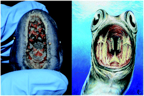
Mineralized tissues such as bones and teeth form the vast majority of what is known of the vertebrate fossil record, whereas non-mineralized tissues are primarily known only from exceptional localities. New chemical techniques have been developed or applied over the last two decades to analyze potential biomarkers for evidence of soft tissues such as keratin, including immunohistochemistry and synchrotron-based chemical analyses among others. These techniques have led to the identification of keratin in fossil feathers and claws by the presence of biologic sulfur residues. Histological sections of Permian and Ordovician aged conodont dental elements are examined for the presence and distribution of soft tissue biomarkers utilizing a suite of spectrometry techniques. Data obtained using energy dispersive X-ray spectrometry consistently show elemental sulfur distributed within the earliest growth stages of the conodont crown as well as in the connected basal body, while X-ray photoelectron spectroscopy analysis supports an organically endogenous origin for at least some of this sulfur. These data suggest that conodont elements, at least in early growth stages, were partly composed of soft tissue, possibly keratin, and were not purely phosphatic. Soft tissue data such as these can have a dramatic effect on our understanding of the early vertebrate fossil record. Incorporating these new data into a phylogenetic analysis suggests conodonts are stem cyclostomes, which is contrary to their current identification as stem gnathostomes.
中文翻译:

光谱技术的新应用揭示了古生代牙形体的系统发育上显着的软组织残留†
脊椎动物化石记录中已知的矿化组织(例如骨骼和牙齿)占绝大多数,而非矿化组织则主要仅在特殊地区才知道。在过去的二十年中,已经开发出或应用了新的化学技术来分析潜在的生物标志物,以寻找诸如角蛋白之类的软组织的证据,包括免疫组织化学和基于同步加速器的化学分析等。这些技术通过生物硫残留物的存在导致了化石羽毛和爪子中角蛋白的鉴定。利用一套光谱技术,检查了二叠纪和奥陶纪的老牙形牙形牙的组织学切片,以了解软组织生物标志物的存在和分布。使用能量色散X射线光谱法获得的数据始终显示出元素硫分布在牙形冠的早期生长阶段以及相连的基体中,而X射线光电子能谱分析至少部分支持了有机内源性来源。硫。这些数据表明,牙形石元素至少在早期生长阶段就部分地由软组织(可能是角蛋白)组成,并且不是纯磷酸盐的。诸如此类的软组织数据可能会对我们对早期脊椎动物化石记录的理解产生巨大影响。将这些新数据纳入系统发育分析表明,牙形石是茎环茎,这与它们目前被鉴定为茎角茎的方法相反。
更新日期:2018-05-08
中文翻译:

光谱技术的新应用揭示了古生代牙形体的系统发育上显着的软组织残留†
脊椎动物化石记录中已知的矿化组织(例如骨骼和牙齿)占绝大多数,而非矿化组织则主要仅在特殊地区才知道。在过去的二十年中,已经开发出或应用了新的化学技术来分析潜在的生物标志物,以寻找诸如角蛋白之类的软组织的证据,包括免疫组织化学和基于同步加速器的化学分析等。这些技术通过生物硫残留物的存在导致了化石羽毛和爪子中角蛋白的鉴定。利用一套光谱技术,检查了二叠纪和奥陶纪的老牙形牙形牙的组织学切片,以了解软组织生物标志物的存在和分布。使用能量色散X射线光谱法获得的数据始终显示出元素硫分布在牙形冠的早期生长阶段以及相连的基体中,而X射线光电子能谱分析至少部分支持了有机内源性来源。硫。这些数据表明,牙形石元素至少在早期生长阶段就部分地由软组织(可能是角蛋白)组成,并且不是纯磷酸盐的。诸如此类的软组织数据可能会对我们对早期脊椎动物化石记录的理解产生巨大影响。将这些新数据纳入系统发育分析表明,牙形石是茎环茎,这与它们目前被鉴定为茎角茎的方法相反。


 京公网安备 11010802027423号