当前位置:
X-MOL 学术
›
Biomater. Sci.
›
论文详情
Our official English website, www.x-mol.net, welcomes your
feedback! (Note: you will need to create a separate account there.)
Cancer cell membrane-coated magnetic nanoparticles for MR/NIR fluorescence dual-modal imaging and photodynamic therapy†
Biomaterials Science ( IF 5.8 ) Pub Date : 2018-05-08 00:00:00 , DOI: 10.1039/c8bm00343b Jiong Li 1, 2, 3, 4, 5 , Xuandong Wang 3, 4, 5, 6, 7 , Dongye Zheng 1, 2, 3, 4, 5 , Xinyi Lin 4, 8, 9, 10, 11 , Zuwu Wei 4, 8, 9, 10, 11 , Da Zhang 4, 8, 9, 10, 11 , Zhuanfang Li 4, 12, 13, 14 , Yun Zhang 3, 4, 5, 6, 7 , Ming Wu 1, 2, 3, 4, 8 , Xiaolong Liu 1, 2, 3, 4, 8
Biomaterials Science ( IF 5.8 ) Pub Date : 2018-05-08 00:00:00 , DOI: 10.1039/c8bm00343b Jiong Li 1, 2, 3, 4, 5 , Xuandong Wang 3, 4, 5, 6, 7 , Dongye Zheng 1, 2, 3, 4, 5 , Xinyi Lin 4, 8, 9, 10, 11 , Zuwu Wei 4, 8, 9, 10, 11 , Da Zhang 4, 8, 9, 10, 11 , Zhuanfang Li 4, 12, 13, 14 , Yun Zhang 3, 4, 5, 6, 7 , Ming Wu 1, 2, 3, 4, 8 , Xiaolong Liu 1, 2, 3, 4, 8
Affiliation
|
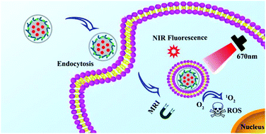
Theranostic nanoprobes integrated with dual-modal imaging and therapeutic functions, such as photodynamic therapy (PDT), have exhibited significant potency in cancer treatments due to their high imaging accuracy and non-invasive advantages for cancer elimination. However, biocompatibility and highly efficient accumulation of these nanoprobes in tumor are still unsatisfactory for clinical application. In this study, a photosensitizer -loaded magnetic nanobead with surface further coated with a layer of cancer cell membrane (SSAP-Ce6@CCM) was designed to improve the biocompatibility and cellular uptake and ultimately achieve enhanced MR/NIR fluorescence imaging and PDT efficacy. Compared with similar nanobeads without CCM coating, SSAP-Ce6@CCM showed significantly enhanced cellular uptake, as evidenced by Prussian blue staining, confocal laser scanning microscopy (CLSM) and flow cytometric analysis. Consequently, SSAP-Ce6@CCM displayed a more distinct MR/NIR imaging ability and more obvious photo-cytotoxicity towards cancer cells under 670 nm laser irradiation. Furthermore, the enhanced PDT effect benefited from the surface coating of cancer cell membrane was demonstrated in SMMC-7721 tumor-bearing mice through tumor growth observation and tumor tissue pathological examination. Therefore, this CCM-disguised nanobead that integrated the abilities of MR/NIR fluorescence dual-modal imaging and photodynamic therapy might be a promising theranostic platform for tumor treatment.
中文翻译:

用于MR / NIR荧光双峰成像和光动力疗法的癌细胞膜涂层磁性纳米粒子†
与双模式成像和治疗功能(例如光动力疗法(PDT))集成的神经治疗学纳米探针由于其高成像精度和消除癌症的非侵入性优势,在癌症治疗中表现出了显着的潜力。然而,这些纳米探针在肿瘤中的生物相容性和高效积累对于临床应用仍不令人满意。在这项研究中,负载光敏剂的磁性纳米珠表面进一步覆盖有一层癌细胞膜(SSAP-Ce6 @ CCM),旨在提高生物相容性和细胞吸收并最终实现增强的MR / NIR荧光成像和PDT功效。与没有CCM涂层的类似纳米珠相比,SSAP-Ce6 @ CCM显示出显着增强的细胞摄取,普鲁士蓝染色证明了这一点,共聚焦激光扫描显微镜(CLSM)和流式细胞仪分析。因此,在670 nm激光照射下,SSAP-Ce6 @ CCM表现出更独特的MR / NIR成像能力和对癌细胞的更明显的光细胞毒性。此外,通过肿瘤生长观察和肿瘤组织病理学检查,在SMMC-7721荷瘤小鼠中证明了受益于癌细胞膜表面涂层的PDT增强作用。因此,这种融合了MR / NIR荧光双模式成像和光动力疗法功能的CCM伪装纳米珠可能是一种有前途的肿瘤治疗学治疗平台。SSAP-Ce6 @ CCM在670 nm激光辐照下显示出更独特的MR / NIR成像能力和对癌细胞的更明显的光细胞毒性。此外,通过肿瘤生长观察和肿瘤组织病理学检查,在SMMC-7721荷瘤小鼠中证明了受益于癌细胞膜表面涂层的PDT增强作用。因此,这种融合了MR / NIR荧光双模式成像和光动力疗法功能的CCM伪装纳米珠可能是一种有前途的肿瘤治疗学治疗平台。SSAP-Ce6 @ CCM在670 nm激光辐照下显示出更独特的MR / NIR成像能力和对癌细胞的更明显的光细胞毒性。此外,通过肿瘤生长观察和肿瘤组织病理学检查,在SMMC-7721荷瘤小鼠中证明了受益于癌细胞膜表面涂层的PDT增强作用。因此,这种融合了MR / NIR荧光双模式成像和光动力疗法功能的CCM伪装纳米珠可能是一种有前途的肿瘤治疗学治疗平台。
更新日期:2018-05-08
中文翻译:
用于MR / NIR荧光双峰成像和光动力疗法的癌细胞膜涂层磁性纳米粒子†
与双模式成像和治疗功能(例如光动力疗法(PDT))集成的神经治疗学纳米探针由于其高成像精度和消除癌症的非侵入性优势,在癌症治疗中表现出了显着的潜力。然而,这些纳米探针在肿瘤中的生物相容性和高效积累对于临床应用仍不令人满意。在这项研究中,负载光敏剂的磁性纳米珠表面进一步覆盖有一层癌细胞膜(SSAP-Ce6 @ CCM),旨在提高生物相容性和细胞吸收并最终实现增强的MR / NIR荧光成像和PDT功效。与没有CCM涂层的类似纳米珠相比,SSAP-Ce6 @ CCM显示出显着增强的细胞摄取,普鲁士蓝染色证明了这一点,共聚焦激光扫描显微镜(CLSM)和流式细胞仪分析。因此,在670 nm激光照射下,SSAP-Ce6 @ CCM表现出更独特的MR / NIR成像能力和对癌细胞的更明显的光细胞毒性。此外,通过肿瘤生长观察和肿瘤组织病理学检查,在SMMC-7721荷瘤小鼠中证明了受益于癌细胞膜表面涂层的PDT增强作用。因此,这种融合了MR / NIR荧光双模式成像和光动力疗法功能的CCM伪装纳米珠可能是一种有前途的肿瘤治疗学治疗平台。SSAP-Ce6 @ CCM在670 nm激光辐照下显示出更独特的MR / NIR成像能力和对癌细胞的更明显的光细胞毒性。此外,通过肿瘤生长观察和肿瘤组织病理学检查,在SMMC-7721荷瘤小鼠中证明了受益于癌细胞膜表面涂层的PDT增强作用。因此,这种融合了MR / NIR荧光双模式成像和光动力疗法功能的CCM伪装纳米珠可能是一种有前途的肿瘤治疗学治疗平台。SSAP-Ce6 @ CCM在670 nm激光辐照下显示出更独特的MR / NIR成像能力和对癌细胞的更明显的光细胞毒性。此外,通过肿瘤生长观察和肿瘤组织病理学检查,在SMMC-7721荷瘤小鼠中证明了受益于癌细胞膜表面涂层的PDT增强作用。因此,这种融合了MR / NIR荧光双模式成像和光动力疗法功能的CCM伪装纳米珠可能是一种有前途的肿瘤治疗学治疗平台。


 京公网安备 11010802027423号