Our official English website, www.x-mol.net, welcomes your
feedback! (Note: you will need to create a separate account there.)
Magnetic interactions and in vitro study of biocompatible hydrocaffeic acid-stabilized Fe–Pt clusters as MRI contrast agents†
RSC Advances ( IF 3.9 ) Pub Date : 2018-04-19 00:00:00 , DOI: 10.1039/c8ra00047f N Kostevšek 1 , S Hudoklin 2 , M E Kreft 2 , I Serša 3 , A Sepe 3 , Z Jagličić 4 , J Vidmar 5 , J Ščančar 5 , S Šturm 1, 6 , S Kobe 1, 6 , K Žužek Rožman 1, 6
RSC Advances ( IF 3.9 ) Pub Date : 2018-04-19 00:00:00 , DOI: 10.1039/c8ra00047f N Kostevšek 1 , S Hudoklin 2 , M E Kreft 2 , I Serša 3 , A Sepe 3 , Z Jagličić 4 , J Vidmar 5 , J Ščančar 5 , S Šturm 1, 6 , S Kobe 1, 6 , K Žužek Rožman 1, 6
Affiliation
|
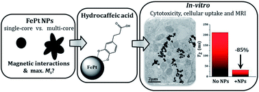
A detailed magnetic study of separated Fe–Pt NPs and Fe–Pt clusters was performed to predict their optimal size and morphology for the maximum saturation magnetization, a factor that is known to influence the performance of a magnetic-resonance-imaging (MRI) contrast agent. Excellent stability and biocompatibility of the nanoparticle suspension was achieved using a novel coating based on hydrocaffeic acid (HCA), which was confirmed with a detailed Fourier-transform infrared spectroscopy (FTIR) study. An in vitro study on a human-bladder papillary urothelial neoplasm RT4 cell line confirmed that HCA-Fe–Pt nanoparticles showed no cytotoxicity, even at a very high concentration (550 μg Fe–Pt per mL), with no delayed cytotoxic effect being detected. This indicates that the HCA coating provides excellent biocompatibility of the nanoparticles, which is a prerequisite for the material to be used as a safe contrast agent for MRI. The cellular uptake and internalization mechanism were studied using ICP-MS and TEM analyses. Furthermore, it was shown that even a very low concentration of Fe–Pt nanoparticles (<10 μg mL−1) in the cells is enough to decrease the T2 relaxation times by 70%. In terms of the MRI imaging, this means a large improvement in the contrast, even at a low nanoparticle concentration and an easier visualization of the tissues containing nanoparticles, proving that HCA-coated Fe–Pt nanoparticles have the potential to be used as an efficient and safe MRI contrast agent.
中文翻译:

生物相容性氢咖啡酸稳定的 Fe-Pt 簇作为 MRI 造影剂的磁相互作用和体外研究†
对分离的 Fe-Pt NPs 和 Fe-Pt 簇进行了详细的磁性研究,以预测其最大饱和磁化强度的最佳尺寸和形态,这是已知会影响磁共振成像 (MRI) 对比度性能的因素代理人。使用基于氢咖啡酸 (HCA) 的新型涂层实现了纳米颗粒悬浮液的出色稳定性和生物相容性,详细的傅里叶变换红外光谱 (FTIR) 研究证实了这一点。体外_对人膀胱乳头状尿路上皮肿瘤 RT4 细胞系的研究证实,HCA-Fe-Pt 纳米粒子即使在非常高的浓度(550 μg Fe-Pt/mL)下也没有显示出细胞毒性,也没有检测到延迟的细胞毒性作用。这表明 HCA 涂层为纳米粒子提供了优异的生物相容性,这是该材料用作 MRI 安全造影剂的先决条件。使用 ICP-MS 和 TEM 分析研究了细胞摄取和内化机制。此外,结果表明,即使细胞中浓度非常低的 Fe-Pt 纳米颗粒 (<10 μg mL -1 ) 也足以降低T 2松弛时间减少 70%。就 MRI 成像而言,这意味着即使在低纳米颗粒浓度下对比度也有很大提高,并且含有纳米颗粒的组织更容易可视化,这证明了 HCA 包覆的 Fe-Pt 纳米颗粒有可能用作有效的和安全的MRI造影剂。
更新日期:2018-04-19
中文翻译:
生物相容性氢咖啡酸稳定的 Fe-Pt 簇作为 MRI 造影剂的磁相互作用和体外研究†
对分离的 Fe-Pt NPs 和 Fe-Pt 簇进行了详细的磁性研究,以预测其最大饱和磁化强度的最佳尺寸和形态,这是已知会影响磁共振成像 (MRI) 对比度性能的因素代理人。使用基于氢咖啡酸 (HCA) 的新型涂层实现了纳米颗粒悬浮液的出色稳定性和生物相容性,详细的傅里叶变换红外光谱 (FTIR) 研究证实了这一点。体外_对人膀胱乳头状尿路上皮肿瘤 RT4 细胞系的研究证实,HCA-Fe-Pt 纳米粒子即使在非常高的浓度(550 μg Fe-Pt/mL)下也没有显示出细胞毒性,也没有检测到延迟的细胞毒性作用。这表明 HCA 涂层为纳米粒子提供了优异的生物相容性,这是该材料用作 MRI 安全造影剂的先决条件。使用 ICP-MS 和 TEM 分析研究了细胞摄取和内化机制。此外,结果表明,即使细胞中浓度非常低的 Fe-Pt 纳米颗粒 (<10 μg mL -1 ) 也足以降低T 2松弛时间减少 70%。就 MRI 成像而言,这意味着即使在低纳米颗粒浓度下对比度也有很大提高,并且含有纳米颗粒的组织更容易可视化,这证明了 HCA 包覆的 Fe-Pt 纳米颗粒有可能用作有效的和安全的MRI造影剂。


 京公网安备 11010802027423号