当前位置:
X-MOL 学术
›
Acc. Chem. Res.
›
论文详情
Our official English website, www.x-mol.net, welcomes your
feedback! (Note: you will need to create a separate account there.)
Optical Probes for Neurobiological Sensing and Imaging
Accounts of Chemical Research ( IF 16.4 ) Pub Date : 2018-04-13 00:00:00 , DOI: 10.1021/acs.accounts.7b00564 Eric H. Kim , Gregory Chin 1 , Guoxin Rong , Kira E. Poskanzer 1 , Heather A. Clark
Accounts of Chemical Research ( IF 16.4 ) Pub Date : 2018-04-13 00:00:00 , DOI: 10.1021/acs.accounts.7b00564 Eric H. Kim , Gregory Chin 1 , Guoxin Rong , Kira E. Poskanzer 1 , Heather A. Clark
Affiliation

|
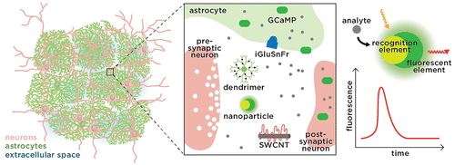
Fluorescent nanosensors and molecular probes are next-generation tools for imaging chemical signaling inside and between cells. Electrophysiology has long been considered the gold standard in elucidating neural dynamics with high temporal resolution and precision, particularly on the single-cell level. However, electrode-based techniques face challenges in illuminating the specific chemicals involved in neural cell activation with adequate spatial information. Measuring chemical dynamics is of fundamental importance to better understand synergistic interactions between neurons as well as interactions between neurons and non-neuronal cells. Over the past decade, significant technological advances in optical probes and imaging methods have enabled entirely new possibilities for studying neural cells and circuits at the chemical level. These optical imaging modalities have shown promise for combining chemical, temporal, and spatial information. This potential makes them ideal candidates to unravel the complex neural interactions at multiple scales in the brain, which could be complemented by traditional electrophysiological methods to obtain a full spatiotemporal picture of neurochemical dynamics. Despite the potential, only a handful of probe candidates have been utilized to provide detailed chemical information in the brain. To date, most live imaging and chemical mapping studies rely on fluorescent molecular indicators to report intracellular calcium (Ca2+) dynamics, which correlates with neuronal activity. Methodological advances for monitoring a full array of chemicals in the brain with improved spatial, temporal, and chemical resolution will thus enable mapping of neurochemical circuits with finer precision.
中文翻译:

用于神经生物学传感和成像的光学探头
荧光纳米传感器和分子探针是用于对细胞内部和细胞之间的化学信号进行成像的下一代工具。长期以来,电生理一直被认为是阐明具有高时间分辨率和精度的神经动力学的金标准,尤其是在单细胞水平上。然而,基于电极的技术在用足够的空间信息阐明神经细胞激活所涉及的特定化学物质方面面临着挑战。测量化学动力学对于更好地理解神经元之间以及神经元与非神经元细胞之间的协同相互作用至关重要。在过去的十年中,光学探针和成像方法的重大技术进步为研究神经细胞和化学级的电路提供了全新的可能性。这些光学成像模态已经显示出结合化学,时间和空间信息的希望。这种潜力使他们成为解开大脑中多个尺度的复杂神经相互作用的理想人选,可以用传统的电生理方法加以补充,以获得神经化学动力学的完整时空图。尽管潜力巨大,但只有少数候选探针可用于在大脑中提供详细的化学信息。迄今为止,大多数实时成像和化学作图研究都依靠荧光分子指标来报告细胞内钙(Ca 可以用传统的电生理方法加以补充,以获得神经化学动力学的完整时空图。尽管潜力巨大,但只有少数候选探针可用于在大脑中提供详细的化学信息。迄今为止,大多数实时成像和化学作图研究都依靠荧光分子指标来报告细胞内钙(Ca 可以用传统的电生理方法加以补充,以获得神经化学动力学的完整时空图。尽管潜力巨大,但只有少数候选探针可用于在大脑中提供详细的化学信息。迄今为止,大多数实时成像和化学作图研究都依靠荧光分子指标来报告细胞内钙(Ca2+)动态,与神经元活动相关。因此,以改进的空间,时间和化学分辨率监测大脑中所有化学物质的方法学进展将使神经化学回路的定位更为精确。
更新日期:2018-04-13
中文翻译:

用于神经生物学传感和成像的光学探头
荧光纳米传感器和分子探针是用于对细胞内部和细胞之间的化学信号进行成像的下一代工具。长期以来,电生理一直被认为是阐明具有高时间分辨率和精度的神经动力学的金标准,尤其是在单细胞水平上。然而,基于电极的技术在用足够的空间信息阐明神经细胞激活所涉及的特定化学物质方面面临着挑战。测量化学动力学对于更好地理解神经元之间以及神经元与非神经元细胞之间的协同相互作用至关重要。在过去的十年中,光学探针和成像方法的重大技术进步为研究神经细胞和化学级的电路提供了全新的可能性。这些光学成像模态已经显示出结合化学,时间和空间信息的希望。这种潜力使他们成为解开大脑中多个尺度的复杂神经相互作用的理想人选,可以用传统的电生理方法加以补充,以获得神经化学动力学的完整时空图。尽管潜力巨大,但只有少数候选探针可用于在大脑中提供详细的化学信息。迄今为止,大多数实时成像和化学作图研究都依靠荧光分子指标来报告细胞内钙(Ca 可以用传统的电生理方法加以补充,以获得神经化学动力学的完整时空图。尽管潜力巨大,但只有少数候选探针可用于在大脑中提供详细的化学信息。迄今为止,大多数实时成像和化学作图研究都依靠荧光分子指标来报告细胞内钙(Ca 可以用传统的电生理方法加以补充,以获得神经化学动力学的完整时空图。尽管潜力巨大,但只有少数候选探针可用于在大脑中提供详细的化学信息。迄今为止,大多数实时成像和化学作图研究都依靠荧光分子指标来报告细胞内钙(Ca2+)动态,与神经元活动相关。因此,以改进的空间,时间和化学分辨率监测大脑中所有化学物质的方法学进展将使神经化学回路的定位更为精确。


 京公网安备 11010802027423号