当前位置:
X-MOL 学术
›
Faraday Discuss.
›
论文详情
Our official English website, www.x-mol.net, welcomes your feedback! (Note: you will need to create a separate account there.)
Functionalised microscale nanoband edge electrode (MNEE) arrays: the systematic quantitative study of hydrogels grown on nanoelectrode biosensor arrays for enhanced sensing in biological media
Faraday Discussions ( IF 3.4 ) Pub Date : 2018-04-12 , DOI: 10.1039/c8fd00063h Andrew Piper 1, 2, 3, 4, 5 , Ben M. Alston 5, 6, 7, 8, 9 , Dave J. Adams 5, 6, 7, 8 , Andrew R. Mount 1, 2, 3, 4, 5
Faraday Discussions ( IF 3.4 ) Pub Date : 2018-04-12 , DOI: 10.1039/c8fd00063h Andrew Piper 1, 2, 3, 4, 5 , Ben M. Alston 5, 6, 7, 8, 9 , Dave J. Adams 5, 6, 7, 8 , Andrew R. Mount 1, 2, 3, 4, 5
Affiliation

|
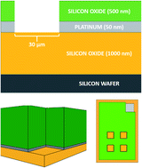
Nanoelectrodes and nanoelectrode arrays show enhanced diffusion and greater faradaic current densities and signal-to-noise ratios compared to macro and microelectrodes, which can lead to enhanced sensing and detection. One example is the microsquare nanoband edge electrode (MNEE) array system, readily formed through microfabrication and whose quantitative response has been established electroanalytically. Hydrogels have been shown to have applications in drug delivery, tissue engineering, and anti-biofouling; some also have the ability to be grown electrochemically. Here, we combine these two emerging technologies to demonstrate the principles of a hydrogel-coated nanoelectrode array biosensor that is resistant to biofouling. We first electrochemically grow and analyze hydrogels on MNEE arrays. The structure of these gels is shown by imaging to be electrochemically controllable, reproducible and structurally hierarchical. This structure is determined by the MNEE array diffusion fields, consistent with the established hydrogel formation reaction, and varies in structural scale from nano (early time, near electrode growth) to micro (for isolated elements in the array) to macro (when there is array overlap) with distance from the electrode, forming a hydrogel mesh of increasing density on progression from solution to electrode. There is also increased hydrogel structural density observed at electrode corners, attributable to enhanced diffusion. The resulting hydrogel structure can be formed on (and is firmly anchored to/through) an established clinically relevant biosensing layer without compromising detection. It is also shown to be capable, through proof-of-principle model protein studies using bovine serum albumin (BSA), of preventing protein biofouling whilst enabling smaller molecules such as DNA to pass through the hydrogel matrix and be sensed. Together, this demonstrates a method for developing reproducible, quantitative electrochemical nanoelectrode biosensors able to sense selectively in real-world sample matrices through the tuning of their interfacial properties.
中文翻译:

功能化的微米级纳米带边缘电极(MNEE)阵列:在纳米电极生物传感器阵列上生长的水凝胶的系统定量研究,用于增强生物介质中的传感
与宏电极和微电极相比,纳米电极和纳米电极阵列显示出增强的扩散和更大的法拉第电流密度以及信噪比,这可以导致增强的感测和检测。一个示例是微正方形纳米带边缘电极(MNEE)阵列系统,该系统很容易通过微细加工形成,并且其定量响应已通过电分析建立。水凝胶已显示出在药物输送,组织工程和抗生物污垢中的应用。有些还具有电化学生长的能力。在这里,我们将这两种新兴技术结合起来,以展示一种具有抗生物结垢功能的水凝胶涂层纳米电极阵列生物传感器的原理。我们首先电化学生长和分析MNEE阵列上的水凝胶。这些凝胶的结构通过成像显示是电化学可控制的,可再现的并且在结构上是分层的。该结构由MNEE阵列扩散场确定,与已建立的水凝胶形成反应一致,并且结构规模从纳米(早期,电极生长附近)到微米(对于阵列中的分离元素)到宏观(存在时)不等。从电极到电极的距离会形成一个水凝胶网,其密度从溶液到电极逐渐增加。在电极角处观察到的水凝胶结构密度也增加了,这归因于扩散的增强。可以在不影响检测的情况下,在已建立的临床相关生物传感层上形成(并牢固地锚定至/穿过)牢固的水凝胶结构。通过使用牛血清白蛋白(BSA)的原理模型蛋白质研究,它还能够防止蛋白质生物积垢,同时使较小的分子(如DNA)穿过水凝胶基质并被感知。总之,这证明了开发可重现的定量电化学纳米电极生物传感器的方法,该传感器能够通过调整其界面特性在现实世界中的样品基质中进行选择性传感。
更新日期:2018-10-10
中文翻译:

功能化的微米级纳米带边缘电极(MNEE)阵列:在纳米电极生物传感器阵列上生长的水凝胶的系统定量研究,用于增强生物介质中的传感
与宏电极和微电极相比,纳米电极和纳米电极阵列显示出增强的扩散和更大的法拉第电流密度以及信噪比,这可以导致增强的感测和检测。一个示例是微正方形纳米带边缘电极(MNEE)阵列系统,该系统很容易通过微细加工形成,并且其定量响应已通过电分析建立。水凝胶已显示出在药物输送,组织工程和抗生物污垢中的应用。有些还具有电化学生长的能力。在这里,我们将这两种新兴技术结合起来,以展示一种具有抗生物结垢功能的水凝胶涂层纳米电极阵列生物传感器的原理。我们首先电化学生长和分析MNEE阵列上的水凝胶。这些凝胶的结构通过成像显示是电化学可控制的,可再现的并且在结构上是分层的。该结构由MNEE阵列扩散场确定,与已建立的水凝胶形成反应一致,并且结构规模从纳米(早期,电极生长附近)到微米(对于阵列中的分离元素)到宏观(存在时)不等。从电极到电极的距离会形成一个水凝胶网,其密度从溶液到电极逐渐增加。在电极角处观察到的水凝胶结构密度也增加了,这归因于扩散的增强。可以在不影响检测的情况下,在已建立的临床相关生物传感层上形成(并牢固地锚定至/穿过)牢固的水凝胶结构。通过使用牛血清白蛋白(BSA)的原理模型蛋白质研究,它还能够防止蛋白质生物积垢,同时使较小的分子(如DNA)穿过水凝胶基质并被感知。总之,这证明了开发可重现的定量电化学纳米电极生物传感器的方法,该传感器能够通过调整其界面特性在现实世界中的样品基质中进行选择性传感。


 京公网安备 11010802027423号