当前位置:
X-MOL 学术
›
Acc. Chem. Res.
›
论文详情
Our official English website, www.x-mol.net, welcomes your
feedback! (Note: you will need to create a separate account there.)
In Situ Cellular Glycan Analysis
Accounts of Chemical Research ( IF 16.4 ) Pub Date : 2018-03-29 00:00:00 , DOI: 10.1021/acs.accounts.7b00617 Yunlong Chen 1 , Lin Ding 1 , Huangxian Ju 1
Accounts of Chemical Research ( IF 16.4 ) Pub Date : 2018-03-29 00:00:00 , DOI: 10.1021/acs.accounts.7b00617 Yunlong Chen 1 , Lin Ding 1 , Huangxian Ju 1
Affiliation

|
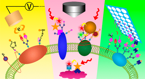
Glycan decorates all mammalian cell surfaces through glycosylation, which is one of the most important post-modifications of proteins. Glycans mediate a wide variety of biological processes, including cell growth and differentiation, cell–cell communication, immune response, pathogen interaction, and intracellular signaling events. Besides, tumor cells aberrantly express distinct sets of glycans, which can indicate different tumor onsets and progression processes. Thus, analysis of cellular glycans may contribute to understanding of glycan-related biological processes and correlation of glycan patterns with disease states for clinical diagnosis and treatment. Although proteomics and glycomics have included great efforts for in vitro study of glycan structures and functions using lysis samples of cells or tissues, they cannot offer real-time qualitative or quantitative information, especially spatial distribution, of glycans on/in intact cells, which is important to the revelation of glycan-related biological events. Moreover, the complex lysis and separation procedures may bring unpredictable loss of glycan information. Focusing on the great urgency for in situ analysis of cellular glycans, our group developed a series of methods for in situ analysis of cellular glycans in the past 10 years. By construction of electrochemical glycan-recognizable probes, glycans on the cell surface can be quantified by direct or competitive electrochemical detection. Using multichannel electrodes or encoded lectin probes, multiple glycans on the cell surface can be dynamically monitored simultaneously. Through design of functional nanoprobes, the cell surface protein-specific glycans and intracellular glycan-related enzymes can be visualized by fluorescence or Raman imaging. Besides, some biological enzymes-based methods have been developed for remodeling or imaging of protein-specific glycans and other types of glycoconjugates, such as gangliosides. Through tracing the changes of glycan expression induced by drugs or gene interference, some glycan-related biological processes have been deduced or proved, demonstrating the reliability and practicability of the developed methods. This Account surveys the key technologies developed in this area, along with the discussion on the shortages of current methodology as well as the possible strategies to overcome those shortages. The future trend in this topic is also discussed. It is expected that this Account can provide a versatile arsenal for chemical and biological researchers to unravel the complex mechanisms involved in glycan-related biological processes and light new beacons in tumor diagnosis and treatment.
中文翻译:

原位细胞聚糖分析
聚糖通过糖基化修饰所有哺乳动物细胞表面,这是蛋白质最重要的后修饰之一。聚糖介导多种生物学过程,包括细胞生长和分化,细胞间通讯,免疫应答,病原体相互作用和细胞内信号事件。此外,肿瘤细胞异常表达不同的聚糖组,这可以指示不同的肿瘤发作和进展过程。因此,对细胞聚糖的分析可能有助于理解聚糖相关的生物学过程,以及聚糖模式与疾病状态之间的相关性,以进行临床诊断和治疗。尽管蛋白质组学和糖组学已经为使用细胞或组织裂解样品进行的聚糖结构和功能的体外研究做出了巨大努力,它们无法提供完整细胞上/内部完整的糖类的实时定性或定量信息,尤其是空间分布,这对于揭示与糖类相关的生物事件很重要。而且,复杂的裂解和分离过程可能带来无法预料的聚糖信息损失。着眼于细胞聚糖原位分析的紧迫性,我们小组在过去的10年中开发了一系列用于细胞聚糖原位分析的方法。通过构建可识别聚糖的电化学探针,可通过直接或竞争性电化学检测对细胞表面的聚糖进行定量。使用多通道电极或编码的凝集素探针,可以同时动态监测细胞表面的多种聚糖。通过功能纳米探针的设计,细胞表面蛋白特异性聚糖和细胞内聚糖相关酶可通过荧光或拉曼成像观察。此外,已经开发了一些基于生物酶的方法,用于蛋白质特异性聚糖和其他类型的糖缀合物(例如神经节苷脂)的重塑或成像。通过追踪药物或基因干扰引起的聚糖表达变化,推论或证明了一些与糖相关的生物学过程,证明了所开发方法的可靠性和实用性。该帐户调查了该领域开发的关键技术,并讨论了当前方法论的不足以及克服这些不足的可能策略。还讨论了该主题的未来趋势。
更新日期:2018-03-29
中文翻译:

原位细胞聚糖分析
聚糖通过糖基化修饰所有哺乳动物细胞表面,这是蛋白质最重要的后修饰之一。聚糖介导多种生物学过程,包括细胞生长和分化,细胞间通讯,免疫应答,病原体相互作用和细胞内信号事件。此外,肿瘤细胞异常表达不同的聚糖组,这可以指示不同的肿瘤发作和进展过程。因此,对细胞聚糖的分析可能有助于理解聚糖相关的生物学过程,以及聚糖模式与疾病状态之间的相关性,以进行临床诊断和治疗。尽管蛋白质组学和糖组学已经为使用细胞或组织裂解样品进行的聚糖结构和功能的体外研究做出了巨大努力,它们无法提供完整细胞上/内部完整的糖类的实时定性或定量信息,尤其是空间分布,这对于揭示与糖类相关的生物事件很重要。而且,复杂的裂解和分离过程可能带来无法预料的聚糖信息损失。着眼于细胞聚糖原位分析的紧迫性,我们小组在过去的10年中开发了一系列用于细胞聚糖原位分析的方法。通过构建可识别聚糖的电化学探针,可通过直接或竞争性电化学检测对细胞表面的聚糖进行定量。使用多通道电极或编码的凝集素探针,可以同时动态监测细胞表面的多种聚糖。通过功能纳米探针的设计,细胞表面蛋白特异性聚糖和细胞内聚糖相关酶可通过荧光或拉曼成像观察。此外,已经开发了一些基于生物酶的方法,用于蛋白质特异性聚糖和其他类型的糖缀合物(例如神经节苷脂)的重塑或成像。通过追踪药物或基因干扰引起的聚糖表达变化,推论或证明了一些与糖相关的生物学过程,证明了所开发方法的可靠性和实用性。该帐户调查了该领域开发的关键技术,并讨论了当前方法论的不足以及克服这些不足的可能策略。还讨论了该主题的未来趋势。


 京公网安备 11010802027423号