当前位置:
X-MOL 学术
›
ACS Chem. Biol.
›
论文详情
Our official English website, www.x-mol.net, welcomes your feedback! (Note: you will need to create a separate account there.)
Unique Fluorescent Imaging Probe for Bacterial Surface Localization and Resistant Enzyme Imaging
ACS Chemical Biology ( IF 4 ) Pub Date : 2018-03-29 00:00:00 , DOI: 10.1021/acschembio.8b00172 Hui Ling Chan 1 , Linna Lyu 1 , Junxin Aw 1 , Wenmin Zhang 1, 2 , Juan Li 2 , Huang-Hao Yang 2 , Hirohito Hayashi 1 , Shunsuke Chiba 1 , Bengang Xing 1, 2
ACS Chemical Biology ( IF 4 ) Pub Date : 2018-03-29 00:00:00 , DOI: 10.1021/acschembio.8b00172 Hui Ling Chan 1 , Linna Lyu 1 , Junxin Aw 1 , Wenmin Zhang 1, 2 , Juan Li 2 , Huang-Hao Yang 2 , Hirohito Hayashi 1 , Shunsuke Chiba 1 , Bengang Xing 1, 2
Affiliation

|
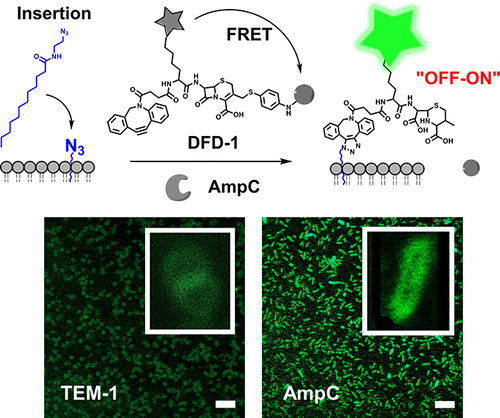
Emergence of antibiotic bacterial resistance has caused serious clinical issues worldwide due to increasingly difficult treatment. Development of a specific approach for selective visualization of resistant bacteria will be highly significant for clinical investigations to promote timely diagnosis and treatment of bacterial infections. In this article, we present an effective method that not only is able to selectively recognize drug resistant AmpC β-lactamases enzyme but, more importantly, is able to interact with bacterial cell wall components, resulting in a desired localization effect on the bacterial surface. A unique and specific enzyme-responsive cephalosporin probe (DFD-1) has been developed for the selective recognition of resistance bacteria AmpC β-lactamase, by employing fluorescence resonance energy transfer with an “off–on” bioimaging. To achieve the desired localization, a lipid–azide conjugate (LA-12) was utilized to facilitate its penetration into the bacterial surface, followed by copper-free click chemistry. This enables the probe DFD-1 to be anchored onto the cell surface. In the presence of AmpC enzymes, the cephalosporin β-lactam ring on DFD-1 will be hydrolyzed, leading to the quencher release, thus generating fluorescence for real-time resistant bacterial screening. More importantly, the bulky dibenzocyclooctyne group in DFD-1 allowed selective recognition toward the AmpC bacterial enzyme instead of its counterpart (e.g., TEM-1 β-lactamase). Both live cell imaging and cell cytometry assays showed the great selectivity of DFD-1 to drug resistant bacterial pathogens containing the AmpC enzyme with significant fluorescence enhancement (∼67-fold). This probe presented promising capability to selectively localize and screen for AmpC resistance bacteria, providing great promise for clinical microbiological applications.
中文翻译:

独特的荧光成像探针,用于细菌表面定位和抗性酶成像
由于越来越难以治疗,抗生素细菌抗性的出现已在世界范围内引起严重的临床问题。开发选择性方法对耐药菌进行选择性可视化的方法,对于促进及时诊断和治疗细菌感染的临床研究将具有重要意义。在本文中,我们提出了一种有效的方法,该方法不仅能够选择性地识别抗药性AmpCβ-内酰胺酶,而且更重要的是能够与细菌细胞壁成分相互作用,从而在细菌表面产生所需的定位作用。独特而独特的酶反应性头孢菌素探针(DFD-1)已经开发出来,通过利用“共振”生物成像技术进行荧光共振能量转移来选择性识别抗药性细菌AmpCβ-内酰胺酶。为了实现所需的定位,利用脂质-叠氮化物共轭物(LA-12)促进其渗透进入细菌表面,然后进行无铜点击化学反应。这使得探针DFD-1可以锚定在细胞表面上。在存在AmpC酶的情况下,DFD-1上的头孢菌素β-内酰胺环将被水解,导致淬灭剂释放,从而产生用于实时耐药细菌筛选的荧光。更重要的是,DFD-1中庞大的二苯并环辛炔基团允许选择性识别AmpC细菌酶而不是其对应物(例如TEM-1β-内酰胺酶)。活细胞成像和细胞流式细胞术检测均显示DFD-1对含有AmpC酶的耐药细菌病原体具有极大的选择性,荧光强度显着提高(约67倍)。该探针具有选择性定位和筛选AmpC抗性细菌的强大潜力,为临床微生物学应用提供了广阔前景。
更新日期:2018-03-29
中文翻译:

独特的荧光成像探针,用于细菌表面定位和抗性酶成像
由于越来越难以治疗,抗生素细菌抗性的出现已在世界范围内引起严重的临床问题。开发选择性方法对耐药菌进行选择性可视化的方法,对于促进及时诊断和治疗细菌感染的临床研究将具有重要意义。在本文中,我们提出了一种有效的方法,该方法不仅能够选择性地识别抗药性AmpCβ-内酰胺酶,而且更重要的是能够与细菌细胞壁成分相互作用,从而在细菌表面产生所需的定位作用。独特而独特的酶反应性头孢菌素探针(DFD-1)已经开发出来,通过利用“共振”生物成像技术进行荧光共振能量转移来选择性识别抗药性细菌AmpCβ-内酰胺酶。为了实现所需的定位,利用脂质-叠氮化物共轭物(LA-12)促进其渗透进入细菌表面,然后进行无铜点击化学反应。这使得探针DFD-1可以锚定在细胞表面上。在存在AmpC酶的情况下,DFD-1上的头孢菌素β-内酰胺环将被水解,导致淬灭剂释放,从而产生用于实时耐药细菌筛选的荧光。更重要的是,DFD-1中庞大的二苯并环辛炔基团允许选择性识别AmpC细菌酶而不是其对应物(例如TEM-1β-内酰胺酶)。活细胞成像和细胞流式细胞术检测均显示DFD-1对含有AmpC酶的耐药细菌病原体具有极大的选择性,荧光强度显着提高(约67倍)。该探针具有选择性定位和筛选AmpC抗性细菌的强大潜力,为临床微生物学应用提供了广阔前景。


 京公网安备 11010802027423号