Our official English website, www.x-mol.net, welcomes your
feedback! (Note: you will need to create a separate account there.)
Precise targeting of cancer metastasis using multi-ligand nanoparticles incorporating four different ligands†
Nanoscale ( IF 5.8 ) Pub Date : 2018-03-29 00:00:00 , DOI: 10.1039/c8nr02513d P. M. Peiris 1, 2, 3, 4 , F. He 1, 2, 3, 4 , G. Covarrubias 1, 2, 3, 4, 5 , S. Raghunathan 1, 2, 3, 4 , O. Turan 1, 2, 3, 4 , M. Lorkowski 1, 2, 3, 4 , B. Gnanasambandam 1, 2, 3, 4 , C. Wu 2, 3, 4, 5, 6 , W. P. Schiemann 2, 3, 4, 6 , E. Karathanasis 1, 2, 3, 4, 5
Nanoscale ( IF 5.8 ) Pub Date : 2018-03-29 00:00:00 , DOI: 10.1039/c8nr02513d P. M. Peiris 1, 2, 3, 4 , F. He 1, 2, 3, 4 , G. Covarrubias 1, 2, 3, 4, 5 , S. Raghunathan 1, 2, 3, 4 , O. Turan 1, 2, 3, 4 , M. Lorkowski 1, 2, 3, 4 , B. Gnanasambandam 1, 2, 3, 4 , C. Wu 2, 3, 4, 5, 6 , W. P. Schiemann 2, 3, 4, 6 , E. Karathanasis 1, 2, 3, 4, 5
Affiliation
|
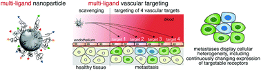
Metastasis displays a highly heterogeneous cellular population with cancer cells continuously evolving. As a result, a single-ligand nanoparticle cannot account for the continuously changing expression of targetable biomarkers over time and space. To effectively direct nanoparticles to metastasis, we developed a multi-ligand nanoparticle by using four different types of ligands on the same nanoparticle that target biomarkers on the endothelium associated with metastatic disease. These vascular targets included αvβ3 integrin, P-selectin, EGFR and fibronectin. Using terminal and in vivo imaging studies, the targeting performance of the multi-ligand nanoparticles was compared to the single-ligand nanoparticle variants. All four single-ligand nanoparticle variants achieved significant targeting of lung metastasis in the 4T1 mouse model of breast cancer metastasis with about 2.5% of the injected dose being deposited into metastasis. A dual-ligand nanoparticle resulted in a nearly 2-fold higher deposition into lung metastases than its single-ligand counterparts. The multi-ligand nanoparticle significantly outperformed its targeting nanoparticle counterparts achieving a deposition of ∼7% of its injected nanoparticles into lung metastases. Using the high sensitivity of radionuclide imaging, PET imaging showed that a multi-ligand nanoparticle labeled with [18F]fluoride was able to precisely target metastatic disease at its very early stage of development in three different animal models of metastatic breast cancer.
中文翻译:

使用包含四个不同配体的多配体纳米颗粒精确靶向癌症转移†
转移表现出高度异质的细胞群体,癌细胞不断发展。结果,单配体纳米颗粒不能解释可靶向生物标志物随时间和空间的不断变化的表达。为了有效地引导纳米颗粒转移,我们通过在同一纳米颗粒上使用四种不同类型的配体来靶向转移性疾病相关内皮上的生物标记物,从而开发了一种多配体纳米颗粒。这些血管目标包括α v β 3整联蛋白,P-选择,EGFR和纤连蛋白。使用终端和体内成像研究中,将多配体纳米颗粒的靶向性能与单配体纳米颗粒变体进行了比较。在乳腺癌转移的4T1小鼠模型中,所有四个单配体纳米粒子变体均实现了针对肺转移的显着靶向,其中约2.5%的注射剂量沉积在了转移中。双配体纳米颗粒比单配体纳米颗粒导致进入肺转移的沉积几乎高2倍。该多配体纳米颗粒的性能明显优于其靶向纳米颗粒,从而将约7%的注射纳米颗粒沉积到了肺转移中。利用放射性核素成像的高灵敏度,PET成像显示标记有[ 18F]氟化物能够在三种不同的转移性乳腺癌动物模型的发展早期准确地靶向转移性疾病。
更新日期:2018-03-29
中文翻译:
使用包含四个不同配体的多配体纳米颗粒精确靶向癌症转移†
转移表现出高度异质的细胞群体,癌细胞不断发展。结果,单配体纳米颗粒不能解释可靶向生物标志物随时间和空间的不断变化的表达。为了有效地引导纳米颗粒转移,我们通过在同一纳米颗粒上使用四种不同类型的配体来靶向转移性疾病相关内皮上的生物标记物,从而开发了一种多配体纳米颗粒。这些血管目标包括α v β 3整联蛋白,P-选择,EGFR和纤连蛋白。使用终端和体内成像研究中,将多配体纳米颗粒的靶向性能与单配体纳米颗粒变体进行了比较。在乳腺癌转移的4T1小鼠模型中,所有四个单配体纳米粒子变体均实现了针对肺转移的显着靶向,其中约2.5%的注射剂量沉积在了转移中。双配体纳米颗粒比单配体纳米颗粒导致进入肺转移的沉积几乎高2倍。该多配体纳米颗粒的性能明显优于其靶向纳米颗粒,从而将约7%的注射纳米颗粒沉积到了肺转移中。利用放射性核素成像的高灵敏度,PET成像显示标记有[ 18F]氟化物能够在三种不同的转移性乳腺癌动物模型的发展早期准确地靶向转移性疾病。


 京公网安备 11010802027423号