当前位置:
X-MOL 学术
›
J. Mater. Process. Tech.
›
论文详情
Our official English website, www.x-mol.net, welcomes your
feedback! (Note: you will need to create a separate account there.)
Micro-voids quantification for damage prediction in warm forging of biocompatible alloys using 3D X-ray CT and RVE approach
Journal of Materials Processing Technology ( IF 6.7 ) Pub Date : 2018-08-01 , DOI: 10.1016/j.jmatprotec.2018.03.020 X.Z. Lu , L.C. Chan
Journal of Materials Processing Technology ( IF 6.7 ) Pub Date : 2018-08-01 , DOI: 10.1016/j.jmatprotec.2018.03.020 X.Z. Lu , L.C. Chan
|
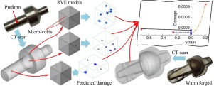
Abstract This study aims to quantify the three-dimensional (3D) micro-voids for damage prediction in warm forging through non-destructive X-ray computed tomography (CT) and RVE approach. Typical biocompatible alloys, i.e., stainless steel 316 L (SS316L) and titanium alloy Ti-6Al-4V, were used as specimen materials in warm-forging a medical implant, i.e., a basal thumb implant. X-ray CT scanning was performed for both the preforms and forged components. Volumetric CT images were then reconstructed and the 3D micro-void distribution and evolution inside the materials were detected and analysed quantitatively. Furthermore, three typical local strain regions, i.e., the small tensile strain region (STSR), small compressive strain region (SCSR) and large compressive strain region (LCSR), were established as the 3D representative volume element (RVE) models for both SS316L and Ti-6Al-4V preforms. The spatial location, size and volume of each micro-void were obtained from defect analysis of the 3D CT images and considered explicitly for subsequent damage prediction. An improved thermo-mechanical coupled micromechanics-based damage (micro-damage) model, which considered the variation of volume fraction of micro-voids (VFMVs), was implemented into finite element (FE) package ABAQUS for localized damage prediction of the RVE models. The damage distributions of the RVE models at different strain levels were visualized and identified. Finally, the localized damage evolutions at both compressive and tensile deformations were predicted and found to match quite well with the findings acquired from CT scanning. Thus, the application of non-destructive X-ray CT measurement of micro-voids, incorporating the RVE approach, was able to play a significant role leading to a more reliable damage prediction in the warm forging process.
中文翻译:

使用 3D X 射线 CT 和 RVE 方法对生物相容性合金温锻损伤预测的微孔量化
摘要 本研究旨在通过无损 X 射线计算机断层扫描 (CT) 和 RVE 方法量化用于温锻损伤预测的三维 (3D) 微孔洞。典型的生物相容性合金,即不锈钢 316 L (SS316L) 和钛合金 Ti-6Al-4V,被用作热锻医疗植入物(即拇指基部植入物)的样本材料。对预制件和锻造部件都进行了 X 射线 CT 扫描。然后重建体积 CT 图像,并定量检测和分析材料内部的 3D 微空隙分布和演变。此外,三个典型的局部应变区,即小拉伸应变区(STSR)、小压缩应变区(SCSR)和大压缩应变区(LCSR),被建立为 SS316L 和 Ti-6Al-4V 预制件的 3D 代表性体积元 (RVE) 模型。每个微孔洞的空间位置、大小和体积是从 3D CT 图像的缺陷分析中获得的,并明确考虑用于后续的损伤预测。一种改进的基于热-机械耦合的微机械损伤(微损伤)模型,该模型考虑了微空隙体积分数 (VFMV) 的变化,被实施到有限元 (FE) 包 ABAQUS 中,用于 RVE 模型的局部损伤预测. RVE 模型在不同应变水平下的损伤分布被可视化和识别。最后,预测了压缩变形和拉伸变形下的局部损伤演变,发现与 CT 扫描获得的结果非常匹配。因此,
更新日期:2018-08-01
中文翻译:
使用 3D X 射线 CT 和 RVE 方法对生物相容性合金温锻损伤预测的微孔量化
摘要 本研究旨在通过无损 X 射线计算机断层扫描 (CT) 和 RVE 方法量化用于温锻损伤预测的三维 (3D) 微孔洞。典型的生物相容性合金,即不锈钢 316 L (SS316L) 和钛合金 Ti-6Al-4V,被用作热锻医疗植入物(即拇指基部植入物)的样本材料。对预制件和锻造部件都进行了 X 射线 CT 扫描。然后重建体积 CT 图像,并定量检测和分析材料内部的 3D 微空隙分布和演变。此外,三个典型的局部应变区,即小拉伸应变区(STSR)、小压缩应变区(SCSR)和大压缩应变区(LCSR),被建立为 SS316L 和 Ti-6Al-4V 预制件的 3D 代表性体积元 (RVE) 模型。每个微孔洞的空间位置、大小和体积是从 3D CT 图像的缺陷分析中获得的,并明确考虑用于后续的损伤预测。一种改进的基于热-机械耦合的微机械损伤(微损伤)模型,该模型考虑了微空隙体积分数 (VFMV) 的变化,被实施到有限元 (FE) 包 ABAQUS 中,用于 RVE 模型的局部损伤预测. RVE 模型在不同应变水平下的损伤分布被可视化和识别。最后,预测了压缩变形和拉伸变形下的局部损伤演变,发现与 CT 扫描获得的结果非常匹配。因此,


 京公网安备 11010802027423号