Our official English website, www.x-mol.net, welcomes your
feedback! (Note: you will need to create a separate account there.)
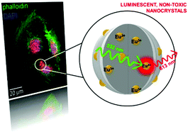
Demonstration of cellular imaging by using luminescent and anti-cytotoxic europium-doped hafnia nanocrystals†
Nanoscale ( IF 5.8 ) Pub Date : 2018-03-27 00:00:00 , DOI: 10.1039/c8nr00724a Irene Villa 1, 2, 3, 4 , Chiara Villa 5, 6, 7, 8, 9 , Angelo Monguzzi 1, 2, 3, 4 , Vladimir Babin 10, 11, 12, 13 , Elena Tervoort 14, 15, 16, 17, 18 , Martin Nikl 10, 11, 12, 13 , Markus Niederberger 14, 15, 16, 17, 18 , Yvan Torrente 5, 6, 7, 8, 9 , Anna Vedda 1, 2, 3, 4 , Alessandro Lauria 14, 15, 16, 17, 18
Nanoscale ( IF 5.8 ) Pub Date : 2018-03-27 00:00:00 , DOI: 10.1039/c8nr00724a Irene Villa 1, 2, 3, 4 , Chiara Villa 5, 6, 7, 8, 9 , Angelo Monguzzi 1, 2, 3, 4 , Vladimir Babin 10, 11, 12, 13 , Elena Tervoort 14, 15, 16, 17, 18 , Martin Nikl 10, 11, 12, 13 , Markus Niederberger 14, 15, 16, 17, 18 , Yvan Torrente 5, 6, 7, 8, 9 , Anna Vedda 1, 2, 3, 4 , Alessandro Lauria 14, 15, 16, 17, 18
Affiliation

|
Luminescent nanoparticles are researched for their potential impact in medical science, but no materials approved for parenteral use have been available so far. To overcome this issue, we demonstrate that Eu3+-doped hafnium dioxide nanocrystals can be used as non-toxic, highly stable probes for cellular optical imaging and as radiosensitive materials for clinical treatment. Furthermore, viability and biocompatibility tests on artificially stressed cell cultures reveal their ability to buffer reactive oxygen species, proposing an anti-cytotoxic feature interesting for biomedical applications.
中文翻译:

通过使用发光和抗细胞毒性的掺euro氧化f纳米晶体演示细胞成像†
研究了发光纳米粒子对医学的潜在影响,但到目前为止,尚无批准用于肠胃外使用的材料。为了克服这个问题,我们证明了Eu 3+掺杂的二氧化f纳米晶体可以用作细胞光学成像的无毒,高稳定性探针,也可以用作临床治疗的放射敏感性材料。此外,在人工应激的细胞培养物中进行的生存能力和生物相容性测试表明,它们具有缓冲活性氧的能力,这为生物医学应用提供了令人感兴趣的抗细胞毒性功能。
更新日期:2018-03-27
中文翻译:

通过使用发光和抗细胞毒性的掺euro氧化f纳米晶体演示细胞成像†
研究了发光纳米粒子对医学的潜在影响,但到目前为止,尚无批准用于肠胃外使用的材料。为了克服这个问题,我们证明了Eu 3+掺杂的二氧化f纳米晶体可以用作细胞光学成像的无毒,高稳定性探针,也可以用作临床治疗的放射敏感性材料。此外,在人工应激的细胞培养物中进行的生存能力和生物相容性测试表明,它们具有缓冲活性氧的能力,这为生物医学应用提供了令人感兴趣的抗细胞毒性功能。


 京公网安备 11010802027423号