Our official English website, www.x-mol.net, welcomes your
feedback! (Note: you will need to create a separate account there.)
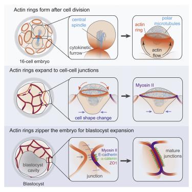
Expanding Actin Rings Zipper the Mouse Embryo for Blastocyst Formation.
Cell ( IF 45.5 ) Pub Date : 2018-Apr-19 , DOI: 10.1016/j.cell.2018.02.035 Jennifer Zenker , Melanie D. White , Maxime Gasnier , Yanina D. Alvarez , Hui Yi Grace Lim , Stephanie Bissiere , Maté Biro , Nicolas Plachta
Cell ( IF 45.5 ) Pub Date : 2018-Apr-19 , DOI: 10.1016/j.cell.2018.02.035 Jennifer Zenker , Melanie D. White , Maxime Gasnier , Yanina D. Alvarez , Hui Yi Grace Lim , Stephanie Bissiere , Maté Biro , Nicolas Plachta
|
Transformation from morula to blastocyst is a defining event of preimplantation embryo development. During this transition, the embryo must establish a paracellular permeability barrier to enable expansion of the blastocyst cavity. Here, using live imaging of mouse embryos, we reveal an actin-zippering mechanism driving this embryo sealing. Preceding blastocyst stage, a cortical F-actin ring assembles at the apical pole of the embryo's outer cells. The ring structure forms when cortical actin flows encounter a network of polar microtubules that exclude F-actin. Unlike stereotypical actin rings, the actin rings of the mouse embryo are not contractile, but instead, they expand to the cell-cell junctions. Here, they couple to the junctions by recruiting and stabilizing adherens and tight junction components. Coupling of the actin rings triggers localized myosin II accumulation, and it initiates a tension-dependent zippering mechanism along the junctions that is required to seal the embryo for blastocyst formation.
中文翻译:

扩大的肌动蛋白环使小鼠胚胎形成胚泡形成拉链。
从桑ula到胚泡的转化是植入前胚胎发育的决定性事件。在此过渡过程中,胚胎必须建立细胞旁通透性屏障,以使胚泡腔扩大。在这里,使用小鼠胚胎的实时成像,我们揭示了驱动该胚胎封闭的肌动蛋白拉链机制。在胚泡期之前,皮质F-肌动蛋白环在胚胎外部细胞的顶极组装。当皮质肌动蛋白流遇到排除F-肌动蛋白的极性微管网络时,形成环结构。与典型的肌动蛋白环不同,小鼠胚胎的肌动蛋白环不收缩,而是扩展到细胞-细胞连接处。在这里,它们通过募集并稳定附着物和紧密的连接组件而耦合到连接处。
更新日期:2018-04-26
中文翻译:
扩大的肌动蛋白环使小鼠胚胎形成胚泡形成拉链。
从桑ula到胚泡的转化是植入前胚胎发育的决定性事件。在此过渡过程中,胚胎必须建立细胞旁通透性屏障,以使胚泡腔扩大。在这里,使用小鼠胚胎的实时成像,我们揭示了驱动该胚胎封闭的肌动蛋白拉链机制。在胚泡期之前,皮质F-肌动蛋白环在胚胎外部细胞的顶极组装。当皮质肌动蛋白流遇到排除F-肌动蛋白的极性微管网络时,形成环结构。与典型的肌动蛋白环不同,小鼠胚胎的肌动蛋白环不收缩,而是扩展到细胞-细胞连接处。在这里,它们通过募集并稳定附着物和紧密的连接组件而耦合到连接处。


 京公网安备 11010802027423号