PLOS ONE ( IF 2.9 ) Pub Date : 2018-03-16 , DOI: 10.1371/journal.pone.0194268 Aloma L. D’Souza , John R. Chevillet , Pejman Ghanouni , Xinrui Yan , Muneesh Tewari , Sanjiv S. Gambhir

|
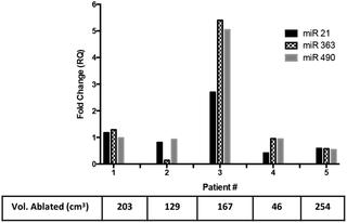
We have previously shown that low frequency ultrasound can release biomarkers from cells into the murine circulation enabling an amplification and localization of the released biomarker that could be used as a blood-based method to detect cancer earlier and monitor therapy. In this study, we further demonstrate that this technique could be used for characterization of tumors and/or identification of cellular masses of unknown origin due to the release of multiple protein and nucleic acid biomarkers in cells in culture, mice and patients. We sonicated colon (LS174T) and prostate (LNCaP) cancer cell lines in culture at a low frequency of 1 MHz and show that there were several-fold changes in multiple protein and microRNA (miRNA) abundance with treatment at various intensities and time. This release was dependent on the duration and intensity of the sonication for both cell lines. Significant increased release in biomarkers was also observed following tumor sonication in living mice bearing subcutaneous LS174T cell line xenografts (for proteins and nucleic acids) and in an experimental LS174T liver tumor model (for proteins only). Finally, we demonstrated this methodology of multiple biomarker release in patients undergoing ablation of uterine fibroids using MR guided high intensity focused ultrasound. Two protein biomarkers significantly increased in the plasma after the ultrasound treatment in 21 samples tested. This proof that ultrasound-amplification method works in soft tissue tumor models together with biomarker multiplexing, could allow for an effective non-invasive method for identification, characterization and localization of incidental lesions, cancer and other disease. Pre-treatment quantification of the biomarkers, allows for individualization of quantitative comparisons. This individualization of normal marker levels in this method allows for specificity of the biomarker-increase to each patient, tumor or organ being studied.
中文翻译:

通过超声释放多种蛋白质和microRNA生物标记物进行肿瘤表征,临床前和临床证据
先前我们已经表明,低频超声可以将生物标志物从细胞释放到鼠循环中,从而可以对释放的生物标志物进行扩增和定位,可以将其用作基于血液的方法,以更早地检测癌症并监测治疗。在这项研究中,我们进一步证明,由于在培养物,小鼠和患者的细胞中释放了多种蛋白质和核酸生物标志物,因此该技术可用于表征肿瘤和/或鉴定未知来源的细胞团。我们在1 MHz的低频下对培养的结肠(LS174T)和前列腺(LNCaP)癌细胞系进行了超声处理,结果表明,在不同强度和时间处理下,多种蛋白质和microRNA(miRNA)丰度都有几倍的变化。该释放取决于两种细胞系的超声处理的持续时间和强度。在携带皮下LS174T细胞系异种移植物(用于蛋白质和核酸)的活小鼠和实验性LS174T肝肿瘤模型(仅用于蛋白质)中进行肿瘤超声处理后,还观察到生物标志物中生物标志物的释放显着增加。最后,我们证明了使用MR引导的高强度聚焦超声在接受子宫肌瘤消融术的患者中释放多种生物标志物的方法。超声处理后的21个样品中,血浆中的两种蛋白质生物标记物显着增加。超声放大方法与生物标志物多路复用一起在软组织肿瘤模型中起作用的这一证据可以为鉴定提供有效的非侵入性方法,偶然病变,癌症和其他疾病的特征和定位。生物标记物的预处理量化可以实现量化比较的个性化。在这种方法中正常标记水平的这种个体化使得生物标记增加对所研究的每个患者,肿瘤或器官的特异性。


 京公网安备 11010802027423号