当前位置:
X-MOL 学术
›
Bioconjugate Chem.
›
论文详情
Our official English website, www.x-mol.net, welcomes your feedback! (Note: you will need to create a separate account there.)
Preoperative Examination and Intraoperative Identification of Hepatocellular Carcinoma Using a Targeted Bimodal Imaging Probe
Bioconjugate Chemistry ( IF 4.7 ) Pub Date : 2018-03-15 00:00:00 , DOI: 10.1021/acs.bioconjchem.8b00161 Yushen Jin 1 , Kun Wang 1 , Jie Tian 1, 2
Bioconjugate Chemistry ( IF 4.7 ) Pub Date : 2018-03-15 00:00:00 , DOI: 10.1021/acs.bioconjchem.8b00161 Yushen Jin 1 , Kun Wang 1 , Jie Tian 1, 2
Affiliation
|
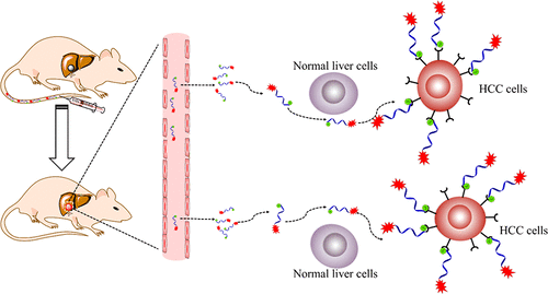
In clinical imaging modalities, MRI is suitable for preoperative examination. Fluorescence imaging has been proposed to improve the detectability of cancer lesions during the operation. However, the specificity and accuracy of the two imaging modalities is limited. To improve the prognosis and survival rate of the patient suffering from hepatocellular carcinoma (HCC), it is very important to develop a specific probe to achieve early detection and precise resection of HCC. We selected HCC targeting peptide SP94 to conjugate with a NIR dye and Gd chelated DOTA to enhance the specificity of different imaging modalities. MRI and fluorescence imaging were used for preoperative examination of the cancer and detecting the tumor in the operation, respectively. MRI and fluorescence signals significantly increased when HCC occurs and the obtained probe shows high uptake in HCC but negligible uptake in the normal liver tissues. The signal-to-background ratio is higher than 2.23 (MRI) and 2.0 (fluorescence imaging) with noninvasive imaging modalities. After the intraperitoneal cavity was surgically exposed, the signal-to-background ratio is higher than 4.6. By combining the high specificity of our probe with the high sensitivity of fluorescence imaging, the microprimary malignancy and micrometastasis foci (diameter <1 mm) can be easily detected after the intraperitoneal cavity is exposed. These results indicated that our probe is conductive to the early detection and precise resection of the HCC and has the potential to improve the diagnosis and treatment of patients with HCC.
中文翻译:

靶向双峰成像探针对肝细胞癌的术前检查和术中鉴定
在临床成像方式中,MRI适用于术前检查。已经提出了荧光成像以改善手术期间癌症病变的可检测性。但是,这两种成像方式的特异性和准确性是有限的。为了提高肝细胞癌(HCC)患者的预后和生存率,开发一种特异性探针以实现肝癌的早期检测和精确切除非常重要。我们选择了HCC靶向肽SP94与NIR染料和Gd螯合的DOTA偶联,以增强不同成像方式的特异性。MRI和荧光成像分别用于术前检查癌症和检测手术中的肿瘤。当发生HCC时,MRI和荧光信号显着增加,并且获得的探针在HCC中显示高摄取,但在正常肝组织中的摄取可忽略不计。使用非侵入性成像方式时,信噪比高于2.23(MRI)和2.0(荧光成像)。腹腔腔手术暴露后,信噪比高于4.6。通过将我们的探针的高特异性与荧光成像的高灵敏度相结合,在腹膜腔暴露后,可以很容易地检测出微原发性恶性肿瘤和微转移灶(直径<1 mm)。这些结果表明,我们的探头有助于肝癌的早期发现和精确切除,并有可能改善肝癌患者的诊断和治疗。
更新日期:2018-03-15
中文翻译:
靶向双峰成像探针对肝细胞癌的术前检查和术中鉴定
在临床成像方式中,MRI适用于术前检查。已经提出了荧光成像以改善手术期间癌症病变的可检测性。但是,这两种成像方式的特异性和准确性是有限的。为了提高肝细胞癌(HCC)患者的预后和生存率,开发一种特异性探针以实现肝癌的早期检测和精确切除非常重要。我们选择了HCC靶向肽SP94与NIR染料和Gd螯合的DOTA偶联,以增强不同成像方式的特异性。MRI和荧光成像分别用于术前检查癌症和检测手术中的肿瘤。当发生HCC时,MRI和荧光信号显着增加,并且获得的探针在HCC中显示高摄取,但在正常肝组织中的摄取可忽略不计。使用非侵入性成像方式时,信噪比高于2.23(MRI)和2.0(荧光成像)。腹腔腔手术暴露后,信噪比高于4.6。通过将我们的探针的高特异性与荧光成像的高灵敏度相结合,在腹膜腔暴露后,可以很容易地检测出微原发性恶性肿瘤和微转移灶(直径<1 mm)。这些结果表明,我们的探头有助于肝癌的早期发现和精确切除,并有可能改善肝癌患者的诊断和治疗。


 京公网安备 11010802027423号