当前位置:
X-MOL 学术
›
JACC Cardiovasc. Imaging
›
论文详情
Our official English website, www.x-mol.net, welcomes your
feedback! (Note: you will need to create a separate account there.)
FDG PET/CT for Early Detection and Localization of Left Ventricular Assist Device Infection: Impact on Patient Management and Outcome.
JACC: Cardiovascular Imaging ( IF 12.8 ) Pub Date : 2018-03-14 , DOI: 10.1016/j.jcmg.2018.01.024 Jongho Kim 1 , Erika D Feller 2 , Wengen Chen 3 , Yuanyuan Liang 4 , Vasken Dilsizian 3
JACC: Cardiovascular Imaging ( IF 12.8 ) Pub Date : 2018-03-14 , DOI: 10.1016/j.jcmg.2018.01.024 Jongho Kim 1 , Erika D Feller 2 , Wengen Chen 3 , Yuanyuan Liang 4 , Vasken Dilsizian 3
Affiliation
|
OBJECTIVES
The feasibility of 18F-fluorodeoxyglucose positron emission tomography/computed tomography (FDG PET/CT) for the diagnosis of left ventricular assist device (LVAD) infection has been demonstrated. Beyond the diagnoses of LVAD infection, the authors hypothesized that the pattern and site of the infection along its various components may significantly impact clinical management and patient outcome.
BACKGROUND
In patients with end-stage heart failure, the clinical use of LVAD for destination therapy is on the rise, accompanied by a higher prevalence of infections and serious complications.
METHODS
FDG PET/CT was performed in 35 heart failure patients with LVAD, 24 with and 11 without clinical suspicion of infection. Microbiology and/or clinical follow-up were used as the final diagnosis standard. Survival rates were compared in patients with and without FDG evidence of infection, and in relation to peripheral (exit wound site or driveline) versus central (cannula or pump) device infection.
RESULTS
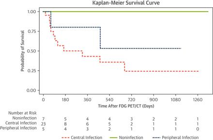
Of 35 patients, 28 (80%) showed metabolic evidence of LVAD infection: 5 limited to the periphery and 23 with extension to the central components of the device. The remaining 7 patients showed no metabolic evidence of infection, which was confirmed by microbiology and clinical follow-up. When CT images were interpreted independently from the FDG PET and clinical information, only 4 of 35 (11%) suggested the possibility of infection. Fourteen of 28 (50%) infected patients died during a mean of 23 months of follow-up after the diagnosis by FDG PET/CT: 12 (86%) with central infection and only 2 with peripheral infection. By contrast, none of the 7 (0%) noninfected patients died (p = 0.03).
CONCLUSIONS
FDG PET/CT is a useful technique for identifying LVAD infection and determining the site and pattern of the infection. The latter has clinical management and patient outcome implications.
中文翻译:

FDG PET / CT用于早期检测和定位左心室辅助装置感染:对患者管理和结果的影响。
目的证明了18F-氟脱氧葡萄糖正电子发射断层扫描/计算机断层扫描(FDG PET / CT)在诊断左心室辅助装置(LVAD)感染中的可行性。除了诊断出LVAD感染外,作者还假设感染的模式和部位及其各个组成部分可能会显着影响临床管理和患者预后。背景技术在患有终末期心力衰竭的患者中,LVAD用于目的地治疗的临床应用正在增加,伴随着较高的感染率和严重的并发症。方法对35例LVAD,24例有11例无临床怀疑感染的心力衰竭患者进行FDG PET / CT检查。微生物学和/或临床随访被用作最终诊断标准。比较有无FDG感染迹象的患者的存活率,并比较外周感染(伤口部位或动力传动系统)与中枢感染(套管或泵)感染的存活率。结果在35例患者中,有28例(80%)显示出LVAD感染的代谢证据:5例局限于外围,23例延伸至设备的中央组件。其余7例患者未显示感染的代谢证据,这已通过微生物学和临床随访得到证实。当CT图像独立于FDG PET和临床信息进行解释时,只有35个患者中有4个(11%)提示可能感染。在FDG PET / CT诊断后平均28个月的随访中,28例感染患者中有14例(50%)死亡:12例(86%)患有中枢性感染,只有2例患有周围性感染。相比之下,7例(0%)未感染患者均未死亡(p = 0.03)。结论FDG PET / CT是鉴定LVAD感染并确定感染部位和方式的有用技术。后者具有临床管理和对患者结果的影响。
更新日期:2019-04-02
中文翻译:
FDG PET / CT用于早期检测和定位左心室辅助装置感染:对患者管理和结果的影响。
目的证明了18F-氟脱氧葡萄糖正电子发射断层扫描/计算机断层扫描(FDG PET / CT)在诊断左心室辅助装置(LVAD)感染中的可行性。除了诊断出LVAD感染外,作者还假设感染的模式和部位及其各个组成部分可能会显着影响临床管理和患者预后。背景技术在患有终末期心力衰竭的患者中,LVAD用于目的地治疗的临床应用正在增加,伴随着较高的感染率和严重的并发症。方法对35例LVAD,24例有11例无临床怀疑感染的心力衰竭患者进行FDG PET / CT检查。微生物学和/或临床随访被用作最终诊断标准。比较有无FDG感染迹象的患者的存活率,并比较外周感染(伤口部位或动力传动系统)与中枢感染(套管或泵)感染的存活率。结果在35例患者中,有28例(80%)显示出LVAD感染的代谢证据:5例局限于外围,23例延伸至设备的中央组件。其余7例患者未显示感染的代谢证据,这已通过微生物学和临床随访得到证实。当CT图像独立于FDG PET和临床信息进行解释时,只有35个患者中有4个(11%)提示可能感染。在FDG PET / CT诊断后平均28个月的随访中,28例感染患者中有14例(50%)死亡:12例(86%)患有中枢性感染,只有2例患有周围性感染。相比之下,7例(0%)未感染患者均未死亡(p = 0.03)。结论FDG PET / CT是鉴定LVAD感染并确定感染部位和方式的有用技术。后者具有临床管理和对患者结果的影响。


 京公网安备 11010802027423号