当前位置:
X-MOL 学术
›
Nanoscale Horiz.
›
论文详情
Our official English website, www.x-mol.net, welcomes your
feedback! (Note: you will need to create a separate account there.)
Nanoscale membrane architecture of healthy and pathological red blood cells†
Nanoscale Horizons ( IF 8.0 ) Pub Date : 2018-03-10 00:00:00 , DOI: 10.1039/c7nh00187h Andra C. Dumitru 1, 2, 3, 4 , Mégane A. Poncin 1, 2, 3, 4 , Louise Conrard 1, 4, 5, 6 , Yves F. Dufrêne 1, 2, 3, 4, 7 , Donatienne Tyteca 1, 4, 5, 6 , David Alsteens 1, 2, 3, 4
Nanoscale Horizons ( IF 8.0 ) Pub Date : 2018-03-10 00:00:00 , DOI: 10.1039/c7nh00187h Andra C. Dumitru 1, 2, 3, 4 , Mégane A. Poncin 1, 2, 3, 4 , Louise Conrard 1, 4, 5, 6 , Yves F. Dufrêne 1, 2, 3, 4, 7 , Donatienne Tyteca 1, 4, 5, 6 , David Alsteens 1, 2, 3, 4
Affiliation
|
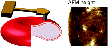
Red blood cells feature remarkable mechanical properties while navigating through microcirculation vessels and during spleen filtration. An unusual combination of plasma membrane and cytoskeleton physical properties allows red blood cells to undergo extensive deformation. Here we used atomic force microscopy multiparametric imaging to probe how cellular organization influences nanoscale and global mechanical properties of cells in both physiological and pathological conditions. Our data obtained in native conditions confirmed that, compared to healthy cells, cells from patients with hereditary spherocytosis are stiffer. Through vertical segmentation of the cell elasticity, we found that healthy and pathological cells display nanoscale architecture with an increasing stiffness along the direction of the applied force. By decoupling the mechanical response of the plasma membrane from its underlying cytoskeleton, we find that both components show altered properties in pathological conditions. Nanoscale multiparametric imaging also revealed lipid domains that exhibit differential mechanical properties than the bulk membrane in both healthy and pathological conditions. Thanks to correlated AFM-fluorescence imaging, we identified submicrometric sphingomyelin-enriched lipid domains of variable stiffness at the red blood cell surface. Our experiments provide novel insights into the interplay between nanoscale organization of red blood cell plasma membrane and their nanomechanical properties. Overall, this work contributes to a better understanding of the complex relationship between cellular nanoscale organization, cellular nanomechanics and how this 3D organization is altered in pathological conditions.
中文翻译:

健康和病理性红细胞的纳米膜结构†
红细胞在微循环血管中和脾脏过滤过程中具有卓越的机械性能。质膜和细胞骨架物理特性的异常组合使红细胞发生广泛的变形。在这里,我们使用原子力显微镜多参数成像技术来探究细胞组织如何在生理和病理条件下影响细胞的纳米级和整体机械特性。我们在自然条件下获得的数据证实,与健康细胞相比,遗传性球囊炎患者的细胞更坚硬。通过细胞弹性的垂直分割,我们发现健康的和病理的细胞显示出纳米级结构,并且沿作用力的方向具有增加的刚度。通过将质膜的机械反应与其潜在的细胞骨架解耦,我们发现这两种成分在病理情况下均显示出改变的特性。纳米级多参数成像还揭示了在健康和病理条件下脂质结构域都比体膜具有不同的机械性能。多亏了相关的原子力显微镜荧光成像,我们在红细胞表面确定了亚微米级鞘磷脂富集的可变硬度的脂质结构域。我们的实验为红细胞质膜的纳米级组织与其纳米机械性能之间的相互作用提供了新颖的见解。总体而言,这项工作有助于更好地了解细胞纳米级组织之间的复杂关系,
更新日期:2018-03-10
中文翻译:
健康和病理性红细胞的纳米膜结构†
红细胞在微循环血管中和脾脏过滤过程中具有卓越的机械性能。质膜和细胞骨架物理特性的异常组合使红细胞发生广泛的变形。在这里,我们使用原子力显微镜多参数成像技术来探究细胞组织如何在生理和病理条件下影响细胞的纳米级和整体机械特性。我们在自然条件下获得的数据证实,与健康细胞相比,遗传性球囊炎患者的细胞更坚硬。通过细胞弹性的垂直分割,我们发现健康的和病理的细胞显示出纳米级结构,并且沿作用力的方向具有增加的刚度。通过将质膜的机械反应与其潜在的细胞骨架解耦,我们发现这两种成分在病理情况下均显示出改变的特性。纳米级多参数成像还揭示了在健康和病理条件下脂质结构域都比体膜具有不同的机械性能。多亏了相关的原子力显微镜荧光成像,我们在红细胞表面确定了亚微米级鞘磷脂富集的可变硬度的脂质结构域。我们的实验为红细胞质膜的纳米级组织与其纳米机械性能之间的相互作用提供了新颖的见解。总体而言,这项工作有助于更好地了解细胞纳米级组织之间的复杂关系,


 京公网安备 11010802027423号