PLOS Neglected Tropical Diseases ( IF 3.4 ) Pub Date : 2018-03-09 , DOI: 10.1371/journal.pntd.0006341 Norio Kasai , Osamu Kondo , Koichi Suzuki , Yoshinori Aoki , Norihisa Ishii , Masamichi Goto
|
Background
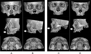
Facial deformation as a sequela of leprosy is caused not only by a saddle nose but also by regression of the maxilla, as well documented in paleopathological observations of excavated skeletal remains of patients with leprosy. However, maxillary changes in living patients have been evaluated only by the subjective visual grading. Here, we attempted to evaluate maxillary bone deformation in patients with leprosy using three-dimensional computed tomography (3D-CT).
Methods
Three-dimensional images centered on the maxilla were reconstructed using multiplanar reconstruction methods in former patients with leprosy (n = 10) and control subjects (n = 5); the anterior-posterior length of the maxilla (MA-P) was then measured. The difference between the MA-P of the patients and those of controls was evaluated after compensating for individual skull size. These findings were also compared with those from previous paleopathological studies.
Findings
Three former patients with lepromatous leprosy showed marked atrophy of the maxilla at the prosthion (-8.6, -11.1 and -17.9 mm) which corresponded with the visual appearance of the maxillary deformity, and these results were consistent with paleopathological findings of excavated skeletal remains. Additionally, the precise bone defects of the maxilla could be individually calculated for accurate reconstructive surgery.
Interpretation
We have successfully illustrated maxillary bone deformities in living patients with leprosy. This study also confirmed the maxillary regression described in paleopathological studies.
中文翻译:
计算机断层扫描技术对麻风病人上颌骨变形的定量评估
背景
作为麻风病后遗症的面部变形不仅由鞍鼻引起,而且由上颌骨的退缩引起,这在麻风病患者挖掘出的骨骼残骸的古病理观察中也有记载。然而,仅通过主观视觉分级评估了活体患者的上颌骨变化。在这里,我们尝试使用三维计算机断层扫描(3D-CT)评估麻风病患者的上颌骨变形。
方法
在以前的麻风病人(n = 10)和对照组(n = 5)中,使用多平面重建方法重建了以上颌骨为中心的三维图像。然后测量上颌的前后长度(MA -P)。补偿个体颅骨大小后,评估患者的M A-P与对照组的M A-P之间的差异。这些发现也与以前的古病理学研究进行了比较。
发现
前三名麻风麻风患者的假体上颌骨明显萎缩(-8.6,-11.1和-17.9 mm),与上颌畸形的外观相符,这些结果与挖掘出的骨骼残留物的古病理结果一致。另外,上颌骨的精确骨缺损可以单独计算以进行精确的重建手术。
解释
我们已经成功地说明了活着的麻风病人的上颌骨畸形。这项研究还证实了古病理学研究中描述的上颌骨退化。


 京公网安备 11010802027423号