PLOS Neglected Tropical Diseases ( IF 3.4 ) Pub Date : 2018-03-09 , DOI: 10.1371/journal.pntd.0006169 Leila M Lopes-Bezerra 1, 2 , Louise A Walker 2 , Gustavo Niño-Vega 3, 4 , Héctor M Mora-Montes 3 , Gabriela W P Neves 1 , Hector Villalobos-Duno 4 , Laura Barreto 4 , Karina Garcia 3 , Bernardo Franco 3 , José A Martínez-Álvarez 3 , Carol A Munro 2 , Neil A R Gow 2
|
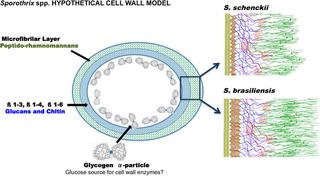
Sporotrichosis is a subcutaneous mycosis caused by pathogenic species of the Sporothrix genus. A new emerging species, Sporothrix brasiliensis, is related to cat-transmitted sporotrichosis and has severe clinical manifestations. The cell wall of pathogenic fungi is a unique structure and impacts directly on the host immune response. We reveal and compare the cell wall structures of Sporothrix schenckii and S. brasiliensis using high-pressure freezing electron microscopy to study the cell wall organization of both species. To analyze the components of the cell wall, we also used infrared and 13C and 1H NMR spectroscopy and the sugar composition was determined by quantitative high-performance anion-exchange chromatography. Our ultrastructural data revealed a bi-layered cell wall structure for both species, including an external microfibrillar layer and an inner electron-dense layer. The inner and outer layers of the S. brasiliensis cell wall were thicker than those of S. schenckii s. str., correlating with an increase in the chitin and rhamnose contents. Moreover, the outer microfibrillar layer of the S. brasiliensis cell wall had longer microfibrils interconnecting yeast cells. Distinct from those of other dimorphic fungi, the cell wall of Sporothrix spp. lacked α-glucan component. Interestingly, glycogen α-particles were identified in the cytoplasm close to the cell wall and the plasma membrane. The cell wall structure as well as the presence of glycogen α-particles varied over time during cell culture. The structural differences observed in the cell wall of these Sporothrix species seemed to impact its uptake by monocyte-derived human macrophages. The data presented here show a unique cell wall structure of S. brasiliensis and S. schenckii during the yeast parasitic phase. A new cell wall model for Sporothrix spp. is therefore proposed that suggests that these fungi molt sheets of intact cell wall layers. This observation may have significant effects on localized and disseminated immunopathology.
中文翻译:
二态真菌病原体申克孢子丝菌和巴西孢子丝菌的细胞壁表现出双层结构和大量完整层的脱落
孢子丝菌病是由孢子丝菌属的病原菌引起的皮下真菌病。一种新出现的物种,巴西孢子丝菌,与猫传播的孢子丝菌病有关,临床表现严重。病原真菌的细胞壁结构独特,直接影响宿主的免疫反应。我们揭示并比较了Sporothrix schenckii和Sporothrix schenckii的细胞壁结构。brasiliensis使用高压冷冻电子显微镜研究这两种物种的细胞壁组织。为了分析细胞壁的成分,我们还使用了红外线和13 C 和1H NMR光谱和糖组成通过定量高效阴离子交换色谱法测定。我们的超微结构数据揭示了这两种物种的双层细胞壁结构,包括外部微纤维层和内部电子致密层。S的内层和外层。brasiliensis细胞壁比S . 申克 str .,与几丁质和鼠李糖含量的增加相关。此外,S的外部微纤维层。brasiliensis细胞壁有更长的微纤维连接酵母细胞。与其他二态真菌不同的是,真菌的细胞壁孢子丝菌属 缺乏α-葡聚糖成分。有趣的是,在靠近细胞壁和质膜的细胞质中发现了糖原 α 粒子。在细胞培养过程中,细胞壁结构以及糖原 α 粒子的存在随时间而变化。在这些孢子丝菌属物种的细胞壁中观察到的结构差异似乎影响了单核细胞衍生的人类巨噬细胞对其的摄取。此处提供的数据显示了S的独特细胞壁结构。巴西和S . schenckii在酵母寄生阶段。一种新的孢子丝菌细胞壁模型spp. 因此提出,这表明这些真菌蜕皮的完整细胞壁层片。这一观察结果可能对局部和播散性免疫病理学产生显着影响。


 京公网安备 11010802027423号