当前位置:
X-MOL 学术
›
The Analyst
›
论文详情
Our official English website, www.x-mol.net, welcomes your
feedback! (Note: you will need to create a separate account there.)
Development and characterization of a scintillating cell imaging dish for radioluminescence microscopy
The Analyst Pub Date : 2018-03-08 , DOI: 10.1039/c8an00106e Debanti Sengupta 1 , Tae Jin Kim 1 , Sepideh Almasi 1 , Stuart Miller 2 , Zsolt Marton 2 , Vivek Nagarkar 2 , Guillem Pratx 1
中文翻译:

用于放射发光显微镜的闪烁细胞成像皿的开发和表征
更新日期:2018-03-08
The Analyst Pub Date : 2018-03-08 , DOI: 10.1039/c8an00106e Debanti Sengupta 1 , Tae Jin Kim 1 , Sepideh Almasi 1 , Stuart Miller 2 , Zsolt Marton 2 , Vivek Nagarkar 2 , Guillem Pratx 1
Affiliation
|
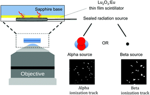
Radioluminescence microscopy is an emerging modality that can be used to image radionuclide probes with micron-scale resolution.
中文翻译:
用于放射发光显微镜的闪烁细胞成像皿的开发和表征
放射发光显微镜是一种新兴的技术,可用于以微米级分辨率对放射性核素探针进行成像。


 京公网安备 11010802027423号