当前位置:
X-MOL 学术
›
Anal. Chem.
›
论文详情
Our official English website, www.x-mol.net, welcomes your
feedback! (Note: you will need to create a separate account there.)
Direct Electrochemiluminescence Imaging of a Single Cell on a Chitosan Film Modified Electrode
Analytical Chemistry ( IF 6.7 ) Pub Date : 2018-03-06 00:00:00 , DOI: 10.1021/acs.analchem.8b00194 Gen Liu 1, 2 , Cheng Ma 2 , Bao-Kang Jin 1 , Zixuan Chen 2 , Jun-Jie Zhu 2
Analytical Chemistry ( IF 6.7 ) Pub Date : 2018-03-06 00:00:00 , DOI: 10.1021/acs.analchem.8b00194 Gen Liu 1, 2 , Cheng Ma 2 , Bao-Kang Jin 1 , Zixuan Chen 2 , Jun-Jie Zhu 2
Affiliation

|
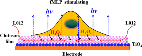
Single-cell imaging is essential for elucidating the biological mechanism of cell function because it accurately reveals the heterogeneity among cells. The electrochemiluminescence (ECL) microscopy technique has been considered a powerful tool to study cells because of its high throughput and zero cellular background light. However, since cells are immobilized on the electrode surface, the steric hindrance and the insulation from the cells make it difficult to obtain a luminous cell ECL image. To solve this problem, direct ECL imaging of a single cell was investigated and achieved on chitosan and nano-TiO2 modified fluoride-doped tin oxide conductive glass (FTO/TiO2/CS). The permeable chitosan film is not only favorable for cell immobilization but also increases the space between the bottom of cells and the electrode; thus, more ECL reagent can exist below the cells compared with the cells on a bare electrode, which guarantees the high sensitivity of quantitative analysis. The modification of nano-TiO2 strengthens the ECL visual signal in luminol solution and effectively improves the signal-to-noise ratio. The light intensity is correlated with the H2O2 concentration on FTO/TiO2/CS, which can be applied to analyze the H2O2 released from cells at the single-cell level. As far as we know, this is the first work to achieve cell ECL imaging without the steric hindrance effect of the cell, and it expands the applications of a modified electrode in visualization study.
中文翻译:

壳聚糖膜修饰电极上单细胞的直接电化学发光成像
单细胞成像对于阐明细胞功能的生物学机制至关重要,因为它可以准确揭示细胞之间的异质性。电化学发光(ECL)显微镜技术因其高通量和零细胞背景光而被认为是研究细胞的强大工具。然而,由于细胞被固定在电极表面上,所以空间位阻和与细胞的绝缘使得难以获得发光细胞ECL图像。为了解决这个问题,研究了在壳聚糖和纳米TiO 2改性氟化物掺杂的氧化锡导电玻璃(FTO / TiO 2)上对单个细胞进行直接ECL成像的研究。/CS)。壳聚糖渗透膜不仅有利于细胞固定,而且增加了细胞底部与电极之间的空间。因此,与裸电极上的细胞相比,细胞下方可以存在更多的ECL试剂,从而保证了定量分析的高灵敏度。纳米TiO 2的改性增强了鲁米诺溶液中的ECL视觉信号,并有效提高了信噪比。光强度与FTO / TiO 2 / CS上的H 2 O 2浓度相关,可用于分析H 2 O 2从单细胞水平的细胞释放。据我们所知,这是在没有细胞空间位阻效应的情况下实现细胞ECL成像的第一项工作,它扩展了修饰电极在可视化研究中的应用。
更新日期:2018-03-06
中文翻译:

壳聚糖膜修饰电极上单细胞的直接电化学发光成像
单细胞成像对于阐明细胞功能的生物学机制至关重要,因为它可以准确揭示细胞之间的异质性。电化学发光(ECL)显微镜技术因其高通量和零细胞背景光而被认为是研究细胞的强大工具。然而,由于细胞被固定在电极表面上,所以空间位阻和与细胞的绝缘使得难以获得发光细胞ECL图像。为了解决这个问题,研究了在壳聚糖和纳米TiO 2改性氟化物掺杂的氧化锡导电玻璃(FTO / TiO 2)上对单个细胞进行直接ECL成像的研究。/CS)。壳聚糖渗透膜不仅有利于细胞固定,而且增加了细胞底部与电极之间的空间。因此,与裸电极上的细胞相比,细胞下方可以存在更多的ECL试剂,从而保证了定量分析的高灵敏度。纳米TiO 2的改性增强了鲁米诺溶液中的ECL视觉信号,并有效提高了信噪比。光强度与FTO / TiO 2 / CS上的H 2 O 2浓度相关,可用于分析H 2 O 2从单细胞水平的细胞释放。据我们所知,这是在没有细胞空间位阻效应的情况下实现细胞ECL成像的第一项工作,它扩展了修饰电极在可视化研究中的应用。


 京公网安备 11010802027423号