当前位置:
X-MOL 学术
›
Anal. Chem.
›
论文详情
Our official English website, www.x-mol.net, welcomes your
feedback! (Note: you will need to create a separate account there.)
Characterization of Tensioned PDMS Membranes for Imaging Cytometry on Microraft Arrays
Analytical Chemistry ( IF 6.7 ) Pub Date : 2018-03-06 00:00:00 , DOI: 10.1021/acs.analchem.8b00176 Matthew DiSalvo 1 , Daniel M. Harris , Saurin Kantesaria 1 , Alexis N. Peña , Jules D. Allbritton-King 1 , Jacqueline H. Cole 1 , Nancy L. Allbritton 1
Analytical Chemistry ( IF 6.7 ) Pub Date : 2018-03-06 00:00:00 , DOI: 10.1021/acs.analchem.8b00176 Matthew DiSalvo 1 , Daniel M. Harris , Saurin Kantesaria 1 , Alexis N. Peña , Jules D. Allbritton-King 1 , Jacqueline H. Cole 1 , Nancy L. Allbritton 1
Affiliation
|
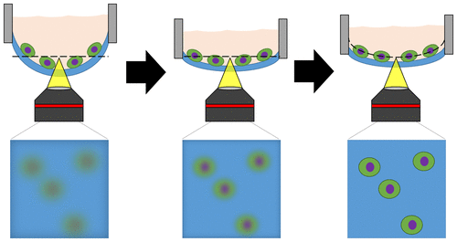
Polydimethylsiloxane (PDMS) membranes can act as sensing elements, barriers, and substrates, yet the low rigidity of the elastomeric membranes can limit their practical use in devices. Microraft arrays rely on a freestanding PDMS membrane as a substrate for cell arrays used in imaging cytometry and cellular isolation. However, the underlying PDMS membrane deforms under the weight of the cell media, making automated analytical microscopy (and thus cytometry and cell isolation) challenging. Here we report the development of microfabrication strategies and physically motivated mathematical modeling of membrane deformation of PDMS microarrays. Microraft arrays were fabricated with mechanical tension stored within the PDMS substrate. These membranes deformed 20× less than that of arrays fabricated using prior methods. Modeling of the deformation of pretensioned arrays using linear membrane theory yielded ≤15% error in predicting the array deflection and predicted the impact of cure temperatures up to 120 °C. A mathematical approach was developed to fit models of microraft shape to sparse real-world shape measurements. Automated imaging of cells on pretensioned microarrays using the focal planes predicted by the model produced high quality fluorescence images of cells, enabling accurate cell area quantification (<4% error) at increased speed (13×) relative to conventional methods. Our microfabrication method and simplified, linear modeling approach is readily applicable to control the deformation of similar membranes in MEMs devices, sensors, and microfluidics.
中文翻译:

张力PDMS膜的表征用于微筏阵列上的细胞计数。
聚二甲基硅氧烷(PDMS)膜可以充当传感元件,屏障和基底,但是弹性体膜的低刚性可能会限制其在设备中的实际使用。Microraft阵列依靠独立的PDMS膜作为成像细胞计数和细胞分离中使用的细胞阵列的底物。但是,下面的PDMS膜在细胞培养基的重量作用下会变形,这使自动分析显微镜检查(以及细胞计数和细胞分离)变得充满挑战。在这里,我们报告了微细加工策略和PDMS微阵列膜变形的物理数学建模的发展。微型筏阵列的机械张力存储在PDMS基板中。这些膜的变形比使用现有方法制造的阵列的变形小20倍。使用线性膜理论对预应力阵列的变形进行建模,在预测阵列挠度和预测高达120°C的固化温度的影响时,产生了≤15%的误差。开发了一种数学方法来拟合微筏形状模型,以稀疏现实世界中的形状测量。使用模型预测的焦平面在预拉伸微阵列上对细胞进行自动成像,可产生高质量的细胞荧光图像,与传统方法相比,能够以提高的速度(13x)进行准确的细胞面积定量(<4%误差)。我们的微细加工方法和简化的线性建模方法很容易应用于控制MEMs设备,传感器和微流体中类似膜的变形。
更新日期:2018-03-06
中文翻译:
张力PDMS膜的表征用于微筏阵列上的细胞计数。
聚二甲基硅氧烷(PDMS)膜可以充当传感元件,屏障和基底,但是弹性体膜的低刚性可能会限制其在设备中的实际使用。Microraft阵列依靠独立的PDMS膜作为成像细胞计数和细胞分离中使用的细胞阵列的底物。但是,下面的PDMS膜在细胞培养基的重量作用下会变形,这使自动分析显微镜检查(以及细胞计数和细胞分离)变得充满挑战。在这里,我们报告了微细加工策略和PDMS微阵列膜变形的物理数学建模的发展。微型筏阵列的机械张力存储在PDMS基板中。这些膜的变形比使用现有方法制造的阵列的变形小20倍。使用线性膜理论对预应力阵列的变形进行建模,在预测阵列挠度和预测高达120°C的固化温度的影响时,产生了≤15%的误差。开发了一种数学方法来拟合微筏形状模型,以稀疏现实世界中的形状测量。使用模型预测的焦平面在预拉伸微阵列上对细胞进行自动成像,可产生高质量的细胞荧光图像,与传统方法相比,能够以提高的速度(13x)进行准确的细胞面积定量(<4%误差)。我们的微细加工方法和简化的线性建模方法很容易应用于控制MEMs设备,传感器和微流体中类似膜的变形。


 京公网安备 11010802027423号