当前位置:
X-MOL 学术
›
Anal. Chem.
›
论文详情
Our official English website, www.x-mol.net, welcomes your
feedback! (Note: you will need to create a separate account there.)
FT-IR Spectroscopic Imaging of Endothelial Cells Response to Tumor Necrosis Factor-α: To Follow Markers of Inflammation Using Standard and High-Magnification Resolution
Analytical Chemistry ( IF 6.7 ) Pub Date : 2018-03-05 00:00:00 , DOI: 10.1021/acs.analchem.7b03089 Ewelina Wiercigroch 1 , Emilia Staniszewska-Slezak 1 , Kinga Szkaradek 2 , Tomasz Wojcik 2 , Yukihiro Ozaki 3 , Malgorzata Baranska 1, 2 , Kamilla Malek 1
Analytical Chemistry ( IF 6.7 ) Pub Date : 2018-03-05 00:00:00 , DOI: 10.1021/acs.analchem.7b03089 Ewelina Wiercigroch 1 , Emilia Staniszewska-Slezak 1 , Kinga Szkaradek 2 , Tomasz Wojcik 2 , Yukihiro Ozaki 3 , Malgorzata Baranska 1, 2 , Kamilla Malek 1
Affiliation

|
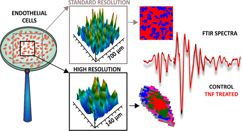
Two endothelial cell lines were selected as models to investigate an effect of incubation with cytokine tumor necrosis factor type α (TNF-α) using Fourier transform infrared (FT-IR) imaging spectroscopy. Both cell lines are often used in laboratories and are typical lung vascular endothelial cells (HMLVEC) derived from the fusion of umbilical vein endothelial cells with lung adenocarcinoma cells (EA.hy926). This study was focused on alteration of spectral changes accompanying inflammation at the cellular level by applying two resolution systems of FT-IR microscopy. The standard approach, with a pixel size of ca. 5.5 μm2, determined the inflammatory state of the whole cell, while a high-magnification resolution (pixel size of ca. 1.1 μm2) provided information at the subcellular level. Importantly, the analysis of IR spectra recorded with different modes produced similar results overall and yielded unambiguous classification of inflamed cells. Generally, the most significant changes in the cells under the influence of TNF-α are related with lipids—their composition and concentration; however, segregation of cells into subcellular compartments provided an additional insight into proteins and nucleic acids related events. The observed spectral alterations are specific for the type of endothelial cell line.
中文翻译:

内皮细胞对肿瘤坏死因子-α反应的FT-IR光谱成像:使用标准和高放大倍数分辨率追踪炎症标记
选择两种内皮细胞系作为模型,以使用傅里叶变换红外(FT-IR)成像光谱技术研究与细胞因子肿瘤坏死因子类型α(TNF-α)孵育的效果。两种细胞系都经常在实验室中使用,并且是典型的肺血管内皮细胞(HMLVEC),其源于脐静脉内皮细胞与肺腺癌细胞(EA.hy926)的融合。这项研究的重点是通过应用FT-IR显微镜的两种分辨率系统,在细胞水平上伴随炎症改变光谱变化。像素大小为ca的标准方法。为5.5μm 2,确定所述全细胞的炎症状态,而高放大倍数的分辨率(大约的1.1微米的像素大小2)提供了亚细胞水平的信息。重要的是,用不同模式记录的红外光谱的分析总体上产生了相似的结果,并且对发炎的细胞进行了明确的分类。通常,在TNF-α的作用下,细胞中最显着的变化与脂质有关-它们的组成和浓度;与脂质有关。然而,将细胞分离到亚细胞区室中提供了对蛋白质和核酸相关事件的进一步了解。观察到的光谱改变对于内皮细胞系的类型是特定的。
更新日期:2018-03-05
中文翻译:

内皮细胞对肿瘤坏死因子-α反应的FT-IR光谱成像:使用标准和高放大倍数分辨率追踪炎症标记
选择两种内皮细胞系作为模型,以使用傅里叶变换红外(FT-IR)成像光谱技术研究与细胞因子肿瘤坏死因子类型α(TNF-α)孵育的效果。两种细胞系都经常在实验室中使用,并且是典型的肺血管内皮细胞(HMLVEC),其源于脐静脉内皮细胞与肺腺癌细胞(EA.hy926)的融合。这项研究的重点是通过应用FT-IR显微镜的两种分辨率系统,在细胞水平上伴随炎症改变光谱变化。像素大小为ca的标准方法。为5.5μm 2,确定所述全细胞的炎症状态,而高放大倍数的分辨率(大约的1.1微米的像素大小2)提供了亚细胞水平的信息。重要的是,用不同模式记录的红外光谱的分析总体上产生了相似的结果,并且对发炎的细胞进行了明确的分类。通常,在TNF-α的作用下,细胞中最显着的变化与脂质有关-它们的组成和浓度;与脂质有关。然而,将细胞分离到亚细胞区室中提供了对蛋白质和核酸相关事件的进一步了解。观察到的光谱改变对于内皮细胞系的类型是特定的。


 京公网安备 11010802027423号