当前位置:
X-MOL 学术
›
J. Am. Chem. Soc.
›
论文详情
Our official English website, www.x-mol.net, welcomes your
feedback! (Note: you will need to create a separate account there.)
Confocal-Raman Microscopy Characterization of Supported Phospholipid Bilayers Deposited on the Interior Surfaces of Chromatographic Silica
Journal of the American Chemical Society ( IF 14.4 ) Pub Date : 2018-02-27 , DOI: 10.1021/jacs.7b13777 David A. Bryce 1 , Jay P. Kitt 1 , Joel M. Harris 1
Journal of the American Chemical Society ( IF 14.4 ) Pub Date : 2018-02-27 , DOI: 10.1021/jacs.7b13777 David A. Bryce 1 , Jay P. Kitt 1 , Joel M. Harris 1
Affiliation

|
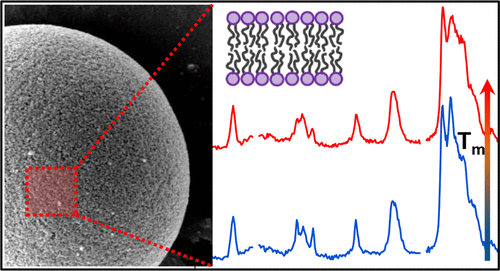
A common approach to exploring the structure and dynamics of biological membranes is through the deposition of model lipid bilayers on planar supports by Langmuir-trough or vesicle-fusion methods. Planar-supported lipid bilayers have been shown to exhibit structure and properties similar to those of lipid-vesicle membranes and are suitable for biosensing applications. Investigations using these planar-membrane models are limited to high-sensitivity methods capable of detecting a small population of molecules at the interface between a planar support and aqueous solution. In this work, we present evidence that supported-lipid bilayers can be deposited by vesicle fusion onto the interior surfaces throughout the wide-pore network of chromatographic silica particles. The thickness of a 1,2-dimyristoyl- sn-glycero-3-phosphocholine (DMPC) film and headgroup spacing are consistent with a single bilayer of DMPC deposited onto the pore surfaces. The high specific surface area of these materials generates phospholipid concentrations easily detected by confocal-Raman microscopy within an individual particle, which allows the structure of these supported bilayers to be investigated. Raman spectra of porous-silica-supported DMPC bilayers are equivalent to spectra of DMPC vesicle membranes, both above and below their melting phase transitions, suggesting comparable phospholipid organization and bilayer structure. These porous-silica-supported model membranes could share benefits that planar-supported lipid bilayers bring to biosensing applications, but in a material that overcomes the limited surface area of a planar support. To test this concept, the potential of these porous-silica-supported lipid bilayers as high-surface-area platforms for label-free Raman-scattering-based protein biosensing is demonstrated with detection of concanavalin A selectively binding to a lipid-immobilized mannose target.
中文翻译:

沉积在色谱二氧化硅内表面的支撑磷脂双层的共聚焦-拉曼显微镜表征
探索生物膜结构和动力学的常用方法是通过 Langmuir-trough 或囊泡融合方法在平面支持物上沉积模型脂质双层。平面支撑的脂双层已被证明具有与脂囊泡膜相似的结构和特性,适用于生物传感应用。使用这些平面膜模型的研究仅限于能够检测平面载体和水溶液之间界面处的少量分子的高灵敏度方法。在这项工作中,我们提供了证据表明支持的脂质双层可以通过囊泡融合沉积到整个色谱二氧化硅颗粒的大孔网络的内表面上。厚度为 1,2-dimyristoyl-sn-glycero-3-phosphocholine (DMPC) 膜和头基间距与沉积在孔表面上的单双层 DMPC 一致。这些材料的高比表面积产生的磷脂浓度很容易通过单个颗粒内的共聚焦拉曼显微镜检测到,从而可以研究这些支持的双层的结构。多孔二氧化硅支撑的 DMPC 双层的拉曼光谱等同于 DMPC 囊泡膜的光谱,高于和低于其熔化相变,表明可比的磷脂组织和双层结构。这些多孔二氧化硅支持的模型膜可以分享平面支持的脂质双层为生物传感应用带来的好处,但在克服平面支持有限表面积的材料中。
更新日期:2018-02-27
中文翻译:

沉积在色谱二氧化硅内表面的支撑磷脂双层的共聚焦-拉曼显微镜表征
探索生物膜结构和动力学的常用方法是通过 Langmuir-trough 或囊泡融合方法在平面支持物上沉积模型脂质双层。平面支撑的脂双层已被证明具有与脂囊泡膜相似的结构和特性,适用于生物传感应用。使用这些平面膜模型的研究仅限于能够检测平面载体和水溶液之间界面处的少量分子的高灵敏度方法。在这项工作中,我们提供了证据表明支持的脂质双层可以通过囊泡融合沉积到整个色谱二氧化硅颗粒的大孔网络的内表面上。厚度为 1,2-dimyristoyl-sn-glycero-3-phosphocholine (DMPC) 膜和头基间距与沉积在孔表面上的单双层 DMPC 一致。这些材料的高比表面积产生的磷脂浓度很容易通过单个颗粒内的共聚焦拉曼显微镜检测到,从而可以研究这些支持的双层的结构。多孔二氧化硅支撑的 DMPC 双层的拉曼光谱等同于 DMPC 囊泡膜的光谱,高于和低于其熔化相变,表明可比的磷脂组织和双层结构。这些多孔二氧化硅支持的模型膜可以分享平面支持的脂质双层为生物传感应用带来的好处,但在克服平面支持有限表面积的材料中。


 京公网安备 11010802027423号