当前位置:
X-MOL 学术
›
Faraday Discuss.
›
论文详情
Our official English website, www.x-mol.net, welcomes your
feedback! (Note: you will need to create a separate account there.)
Investigation of modified nanopore arrays using FIB/SEM tomography
Faraday Discussions ( IF 3.3 ) Pub Date : 2018-02-23 , DOI: 10.1039/c8fd00019k Angelika Holzinger 1, 2, 3, 4 , Gregor Neusser 1, 2, 3, 4 , Benjamin J. J. Austen 5, 6, 7, 8 , Alonso Gamero-Quijano 9, 10, 11, 12, 13 , Grégoire Herzog 9, 10, 11, 12, 13 , Damien W. M. Arrigan 5, 6, 7, 8 , Andreas Ziegler 2, 3, 4, 14 , Paul Walther 2, 3, 4, 14 , Christine Kranz 1, 2, 3, 4
Faraday Discussions ( IF 3.3 ) Pub Date : 2018-02-23 , DOI: 10.1039/c8fd00019k Angelika Holzinger 1, 2, 3, 4 , Gregor Neusser 1, 2, 3, 4 , Benjamin J. J. Austen 5, 6, 7, 8 , Alonso Gamero-Quijano 9, 10, 11, 12, 13 , Grégoire Herzog 9, 10, 11, 12, 13 , Damien W. M. Arrigan 5, 6, 7, 8 , Andreas Ziegler 2, 3, 4, 14 , Paul Walther 2, 3, 4, 14 , Christine Kranz 1, 2, 3, 4
Affiliation
|
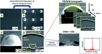
The investigation of electrochemical processes at the interface of two immiscible electrolyte solutions (ITIES) is of great interest for sensing applications, and serves as a surrogate to the study of biological transport phenomena, e.g. ion channels. Alongside e-beam lithography, focused ion beam (FIB) milling is an attractive method to prototype and fabricate nanopore arrays that support nanoITIES. Within this contribution, we explore the capability of FIB/scanning electron microscopy (SEM) tomography to visualize the actual pore structure and interfaces at silica-modified nanoporous membranes. The nanopores were also characterized by atomic force microscopy (AFM) using ultra-sharp AFM probes to determine the pore diameter, and using scanning transmission electron microscopy (STEM) and energy dispersive X-ray (EDX) spectroscopy, providing additional information on the elemental composition of deposits within the pores. Si-rich particles could be identified within the pores as well as at the orifice that had faced the organic electrolyte solution during electrochemical deposition. The prospects of the used techniques for investigating the interface at or within FIB-milled nanopores will be discussed.
中文翻译:

使用FIB / SEM层析成像技术研究修饰的纳米孔阵列
在两种不混溶的电解质溶液(ITIES)的界面上对电化学过程的研究对于传感应用非常重要,并且可以替代对生物传输现象的研究,例如离子通道。除了电子束光刻技术外,聚焦离子束(FIB)铣削是一种有吸引力的方法,可以原型和制造支持nanoITIES的纳米孔阵列。在这项贡献中,我们探索了FIB /扫描电子显微镜(SEM)层析成像的能力,以可视化二氧化硅修饰的纳米多孔膜的实际孔结构和界面。纳米孔的特征还在于使用超锐利AFM探针的原子力显微镜(AFM)来确定孔径,并使用扫描透射电子显微镜(STEM)和能量色散X射线(EDX)光谱,提供有关元素的附加信息。孔内沉积物的组成。可以在孔中以及在电化学沉积过程中面对有机电解质溶液的孔中发现富硅颗粒。将讨论用于研究FIB研磨的纳米孔处或内部的界面的已用技术的前景。
更新日期:2018-10-10
中文翻译:
使用FIB / SEM层析成像技术研究修饰的纳米孔阵列
在两种不混溶的电解质溶液(ITIES)的界面上对电化学过程的研究对于传感应用非常重要,并且可以替代对生物传输现象的研究,例如离子通道。除了电子束光刻技术外,聚焦离子束(FIB)铣削是一种有吸引力的方法,可以原型和制造支持nanoITIES的纳米孔阵列。在这项贡献中,我们探索了FIB /扫描电子显微镜(SEM)层析成像的能力,以可视化二氧化硅修饰的纳米多孔膜的实际孔结构和界面。纳米孔的特征还在于使用超锐利AFM探针的原子力显微镜(AFM)来确定孔径,并使用扫描透射电子显微镜(STEM)和能量色散X射线(EDX)光谱,提供有关元素的附加信息。孔内沉积物的组成。可以在孔中以及在电化学沉积过程中面对有机电解质溶液的孔中发现富硅颗粒。将讨论用于研究FIB研磨的纳米孔处或内部的界面的已用技术的前景。


 京公网安备 11010802027423号