Journal of Controlled Release ( IF 10.8 ) Pub Date : 2018-02-21 , DOI: 10.1016/j.jconrel.2018.02.029 Bongseop Kwak , Yoohwan Lee , Jaehun Lee , Sungwon Lee , Jiseok Lim
|
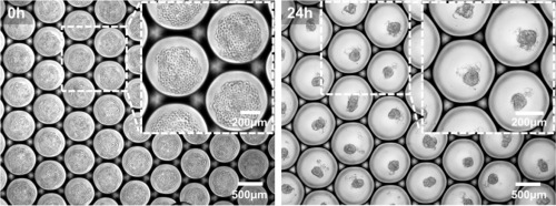
In vivo tumors develop in a three-dimensional manner and have unique and complex characteristics. Physico-biochemical barriers on tumors cause drug resistance and limit drug delivery efficiency. Currently, 2D cancer cell monolayer platforms are frequently used to test the efficiency of new drug materials. However, the monolayer platform generally overestimates drug efficiency because of the absence of physico-biochemical barriers. Many literatures indicated that a 3D tumor spheroid model has very similar characteristics to in vivo tumor models, and studies demonstrated the accurate prediction of drug efficiency using this model. The use of a 3D tumor spheroid model in drug development process remains challenging because of the low generation yield and difficulties in size control. In this study, we developed a droplet-based microfluidic system that can generate cancer cells encapsulated by micro-droplets with very high generation yield (16–20 Hz, 1000 droplets/min). The system can control the number of encapsulated cancer cells in the droplet or diameter of the 3D spheroid model precisely between 50 and 150 μm. Moreover, the formed 3D tumor spheroid model can be cultured for >2 weeks by an additional step of droplet disruption and recollection, and can grow up to 245 μm in diameter.
中文翻译:
使用高通量微流体系统大规模制造均一尺寸的3D肿瘤球体
体内肿瘤以三维方式发展,并具有独特而复杂的特征。肿瘤上的物理生化屏障会导致耐药性并限制药物输送效率。当前,二维癌细胞单层平台经常用于测试新药物材料的效率。但是,由于没有物理生化屏障,单层平台通常会高估药物效率。许多文献表明3D肿瘤球体模型具有与体内非常相似的特征肿瘤模型,并且研究表明使用该模型可以准确预测药物疗效。由于产生产率低且尺寸控制困难,在药物开发过程中使用3D肿瘤球体模型仍然具有挑战性。在这项研究中,我们开发了一种基于液滴的微流控系统,该系统可以产生由微滴包裹的癌细胞,并具有很高的发电量(16–20 Hz,1000滴/分钟)。该系统可以将液滴中的封装癌细胞数或3D球体模型的直径精确控制在50至150μm之间。此外,形成的3D肿瘤球体模型可以通过液滴分裂和回收的额外步骤培养> 2周,并且直径可以增长到245μm。


 京公网安备 11010802027423号